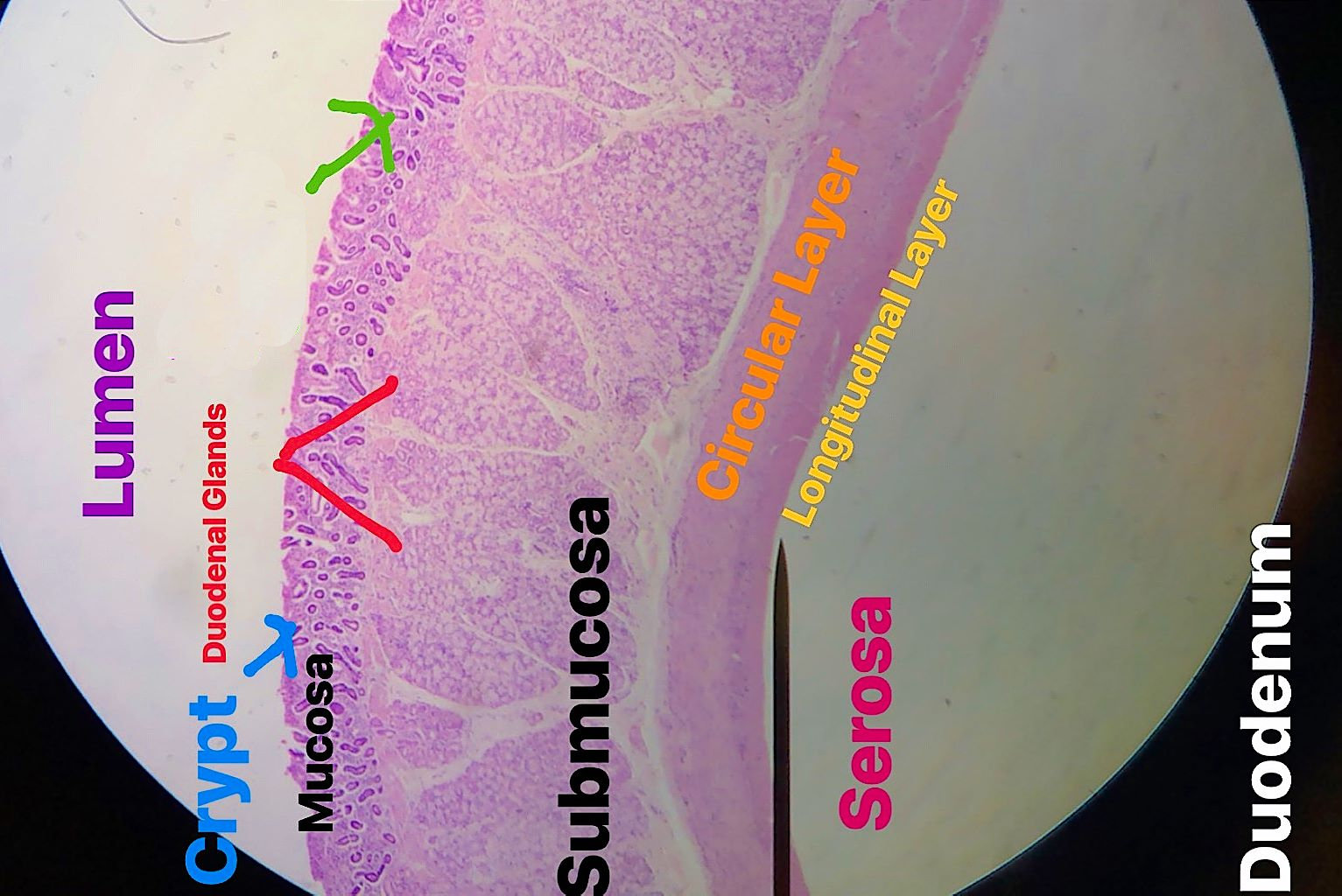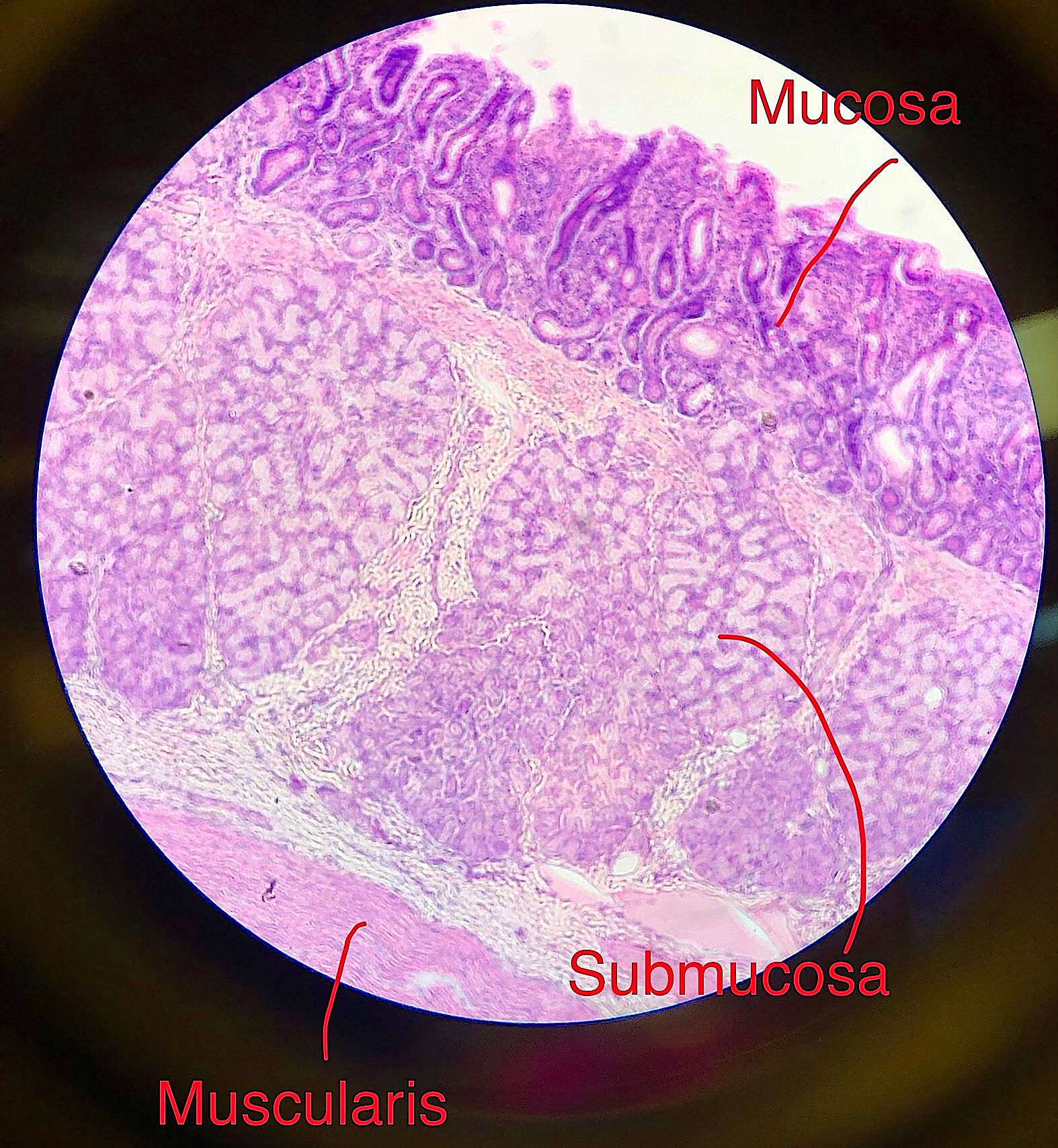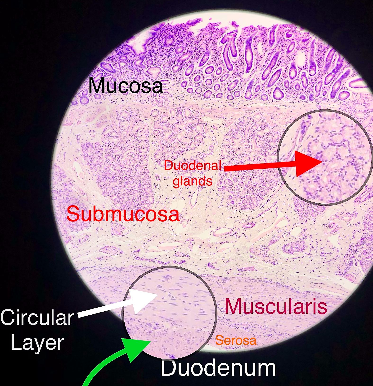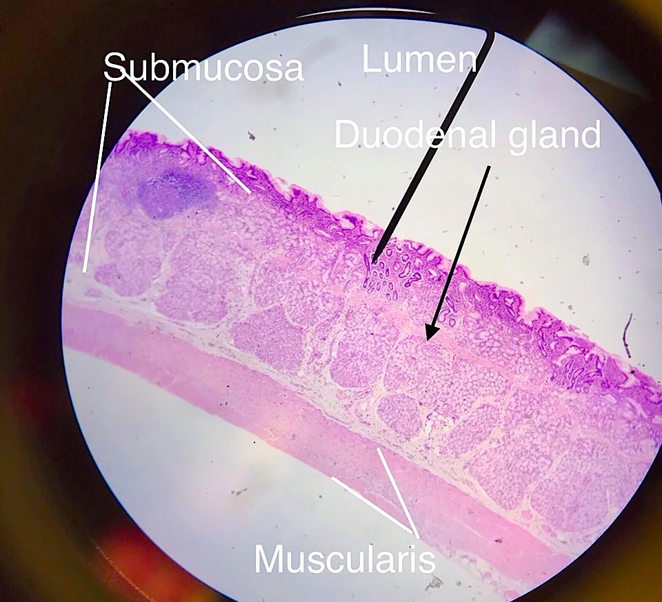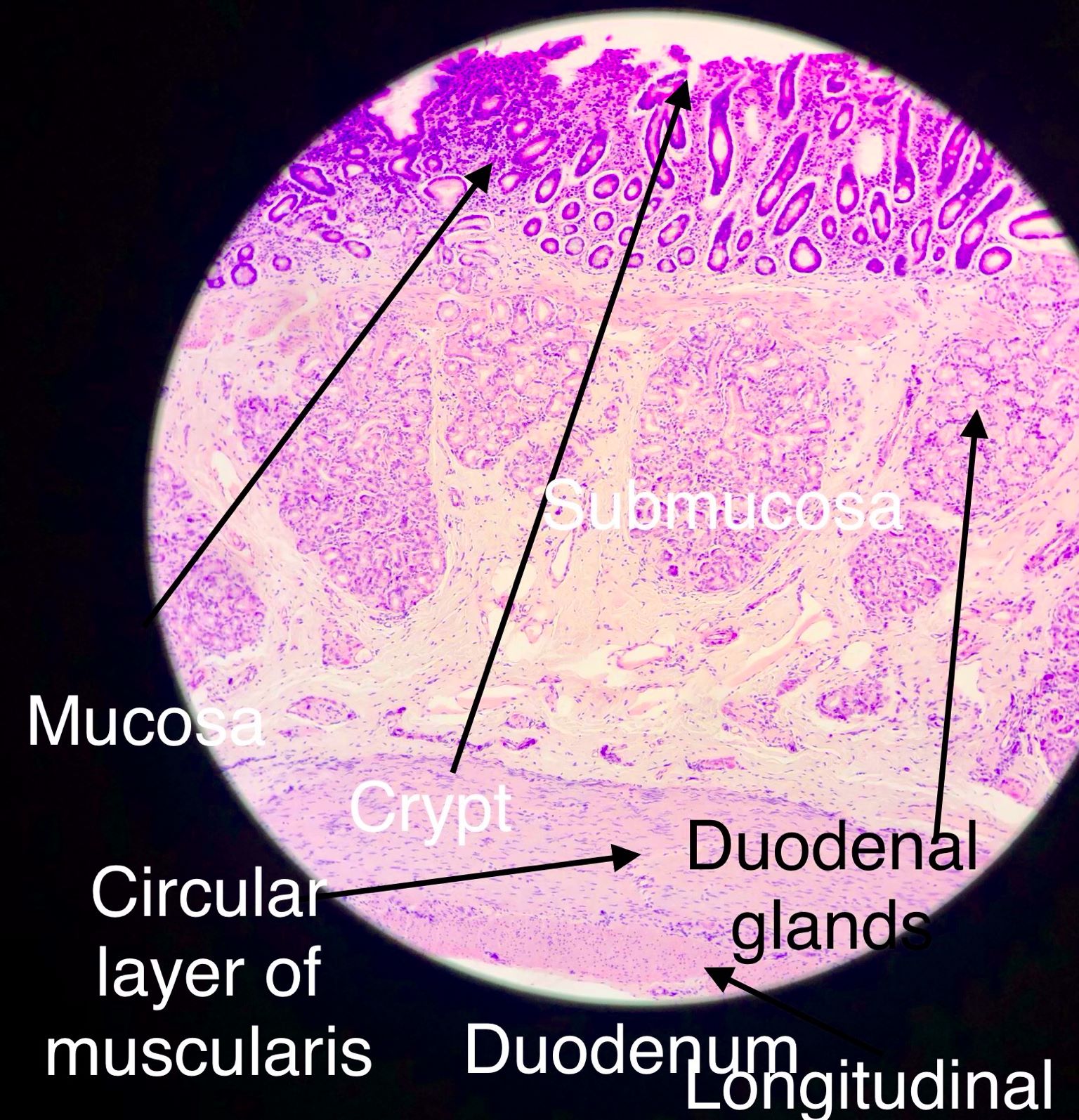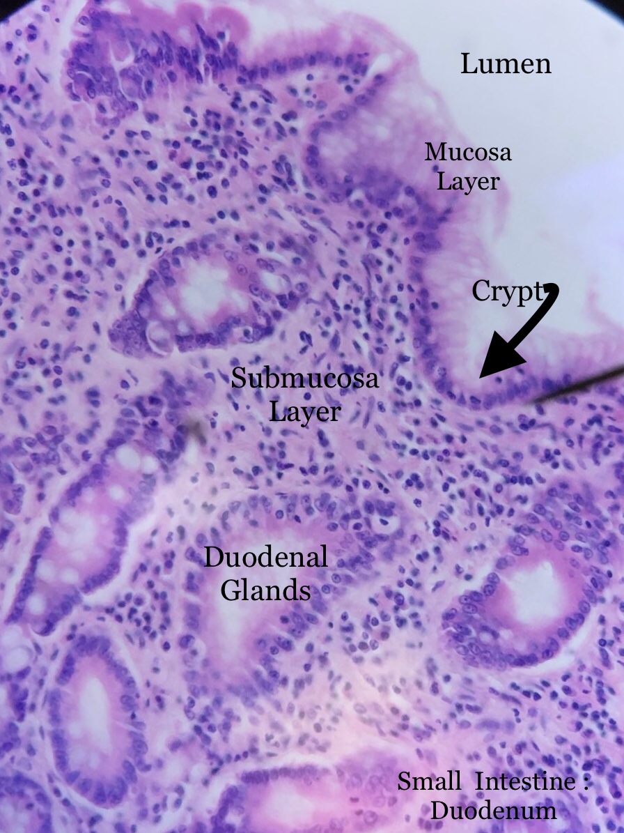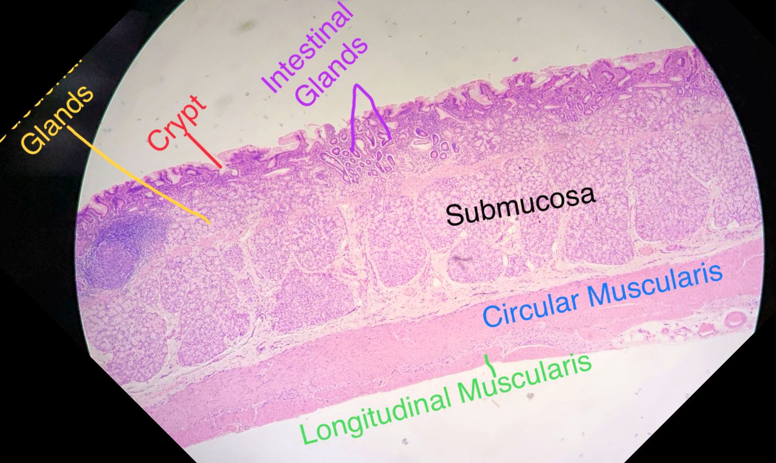
The duodenum, like the rest of the small intestine, has mucosal, submucosal, muscularis, and serosal layers. Furthermore, the muscularis is divided into an inner circular and outer longitudinal layer. The duodenum has very small villi and small crypts at their bases. The submucosa has numerous Brunner's (mucous) gland which is another identifiable feature of this section.
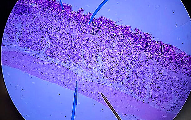
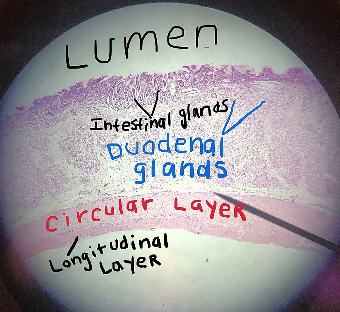
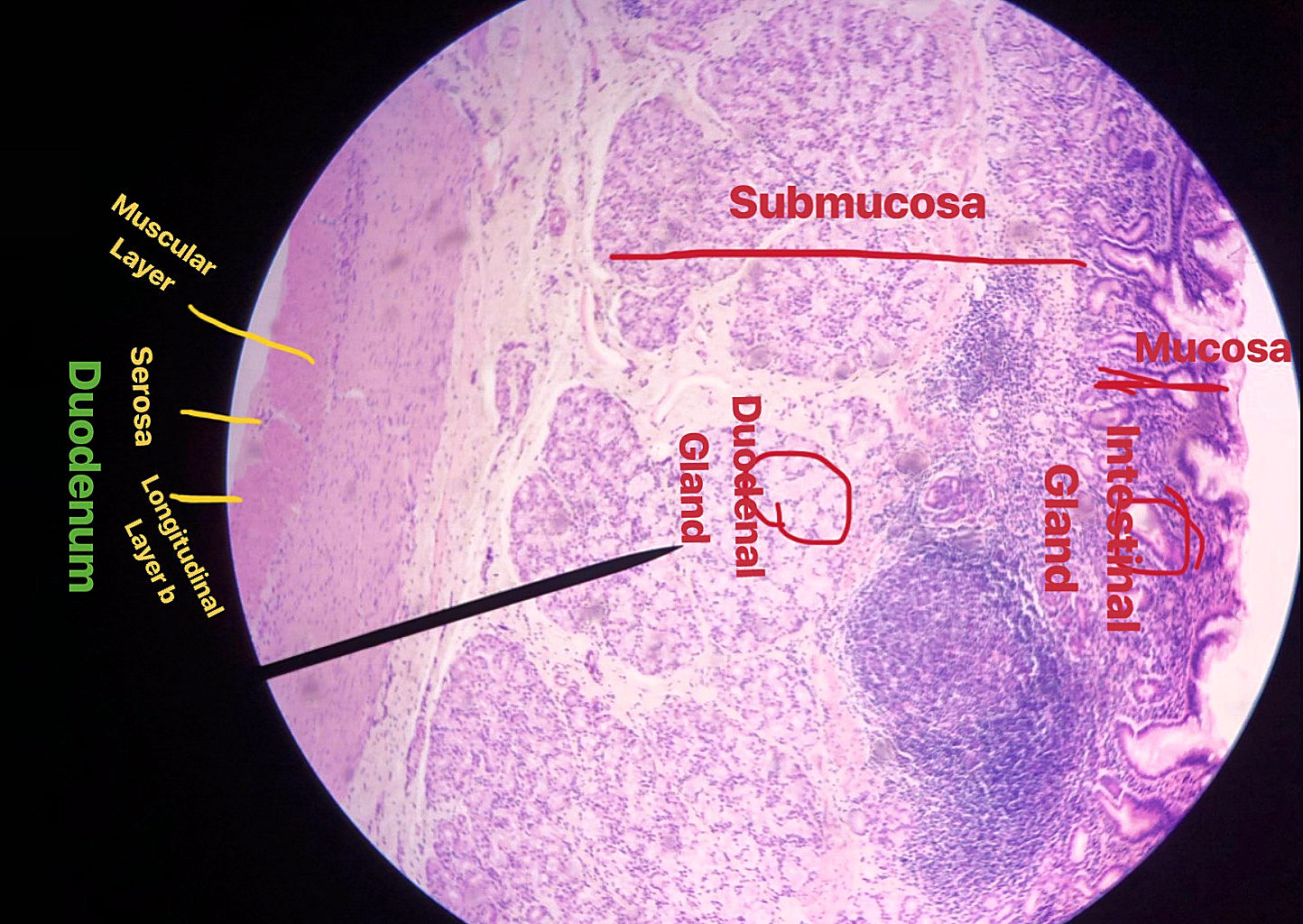
These pictures were taken by bio 139 students in the spring of 2018 and fall of 2019. Scroll through the pictures and compare them with the labeled picture. Select one and draw it. Also see if you can find the 3 pictures with lymph nodules. (Hint, one is the exact same slide as the labeled one but from another student) This model shows what the layers should be also. IT is sort of a combnation of all 3 layers sections.
| Lab Book Image | Student Images |
|---|---|
|
|
|