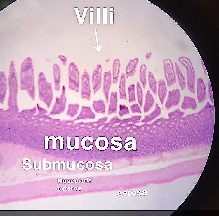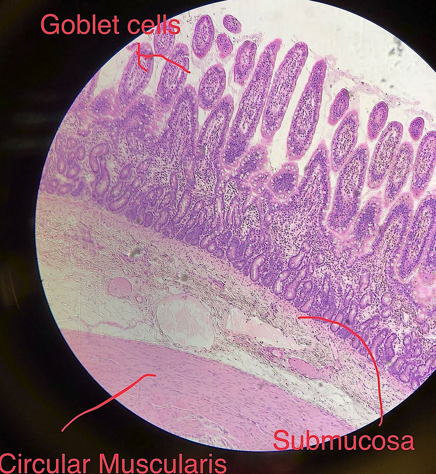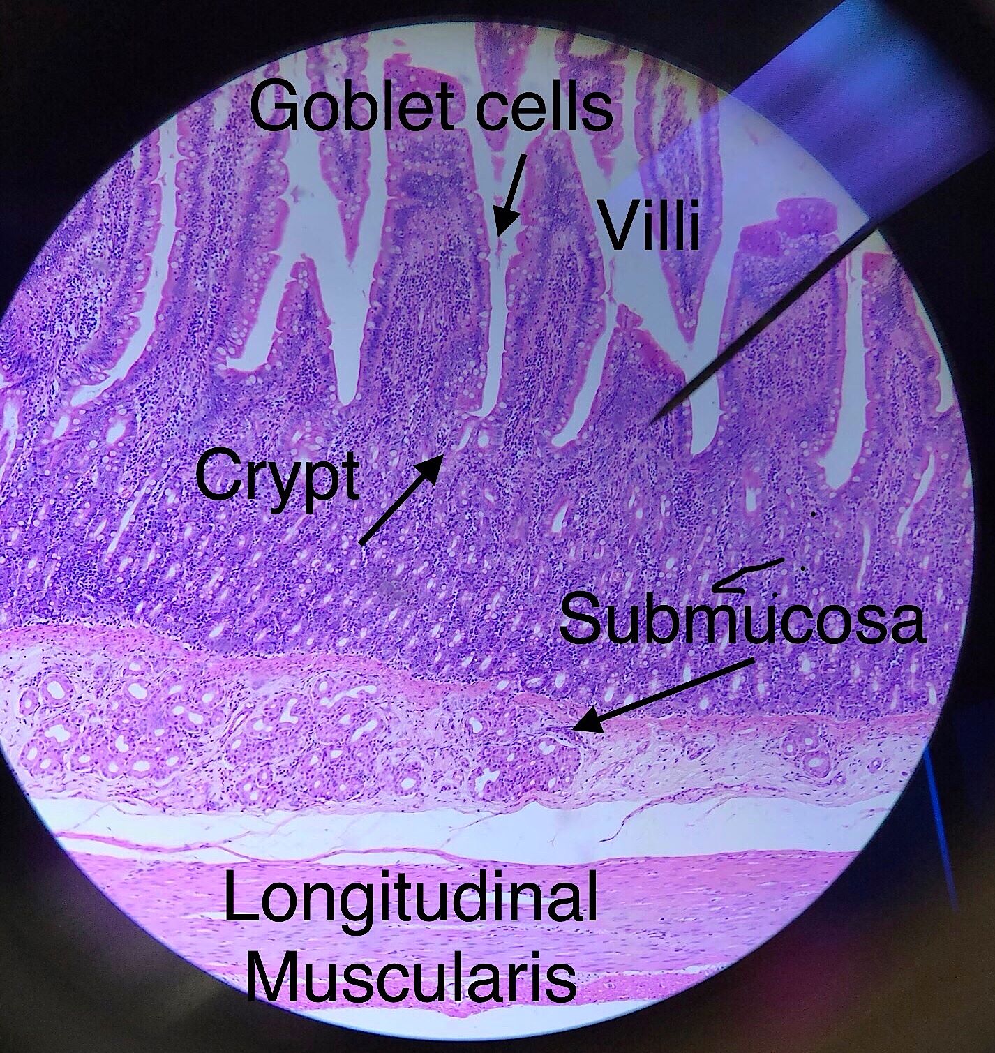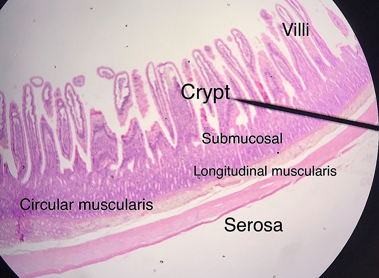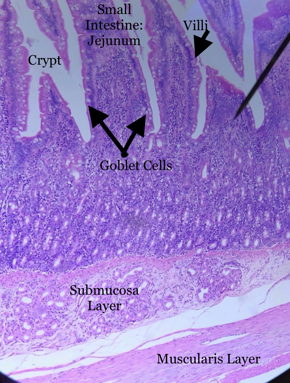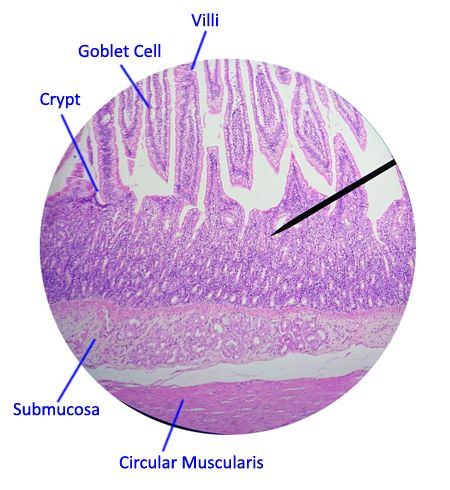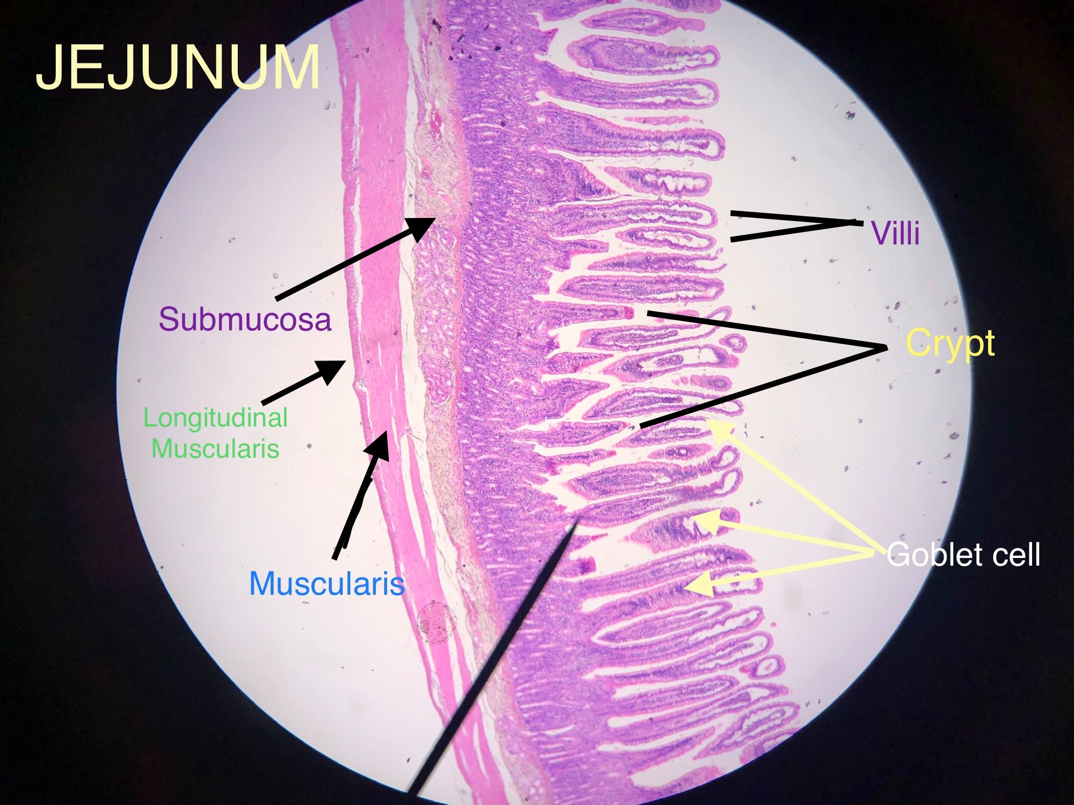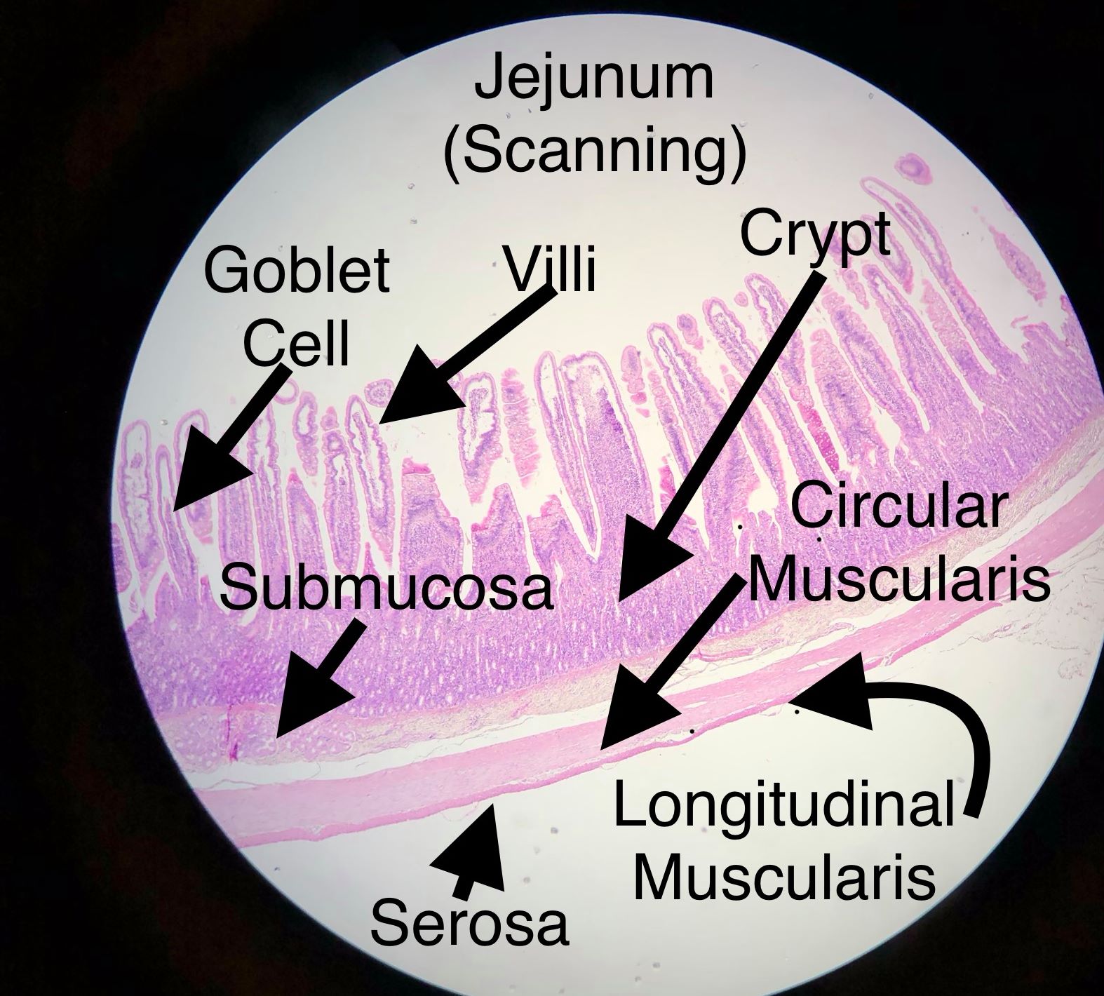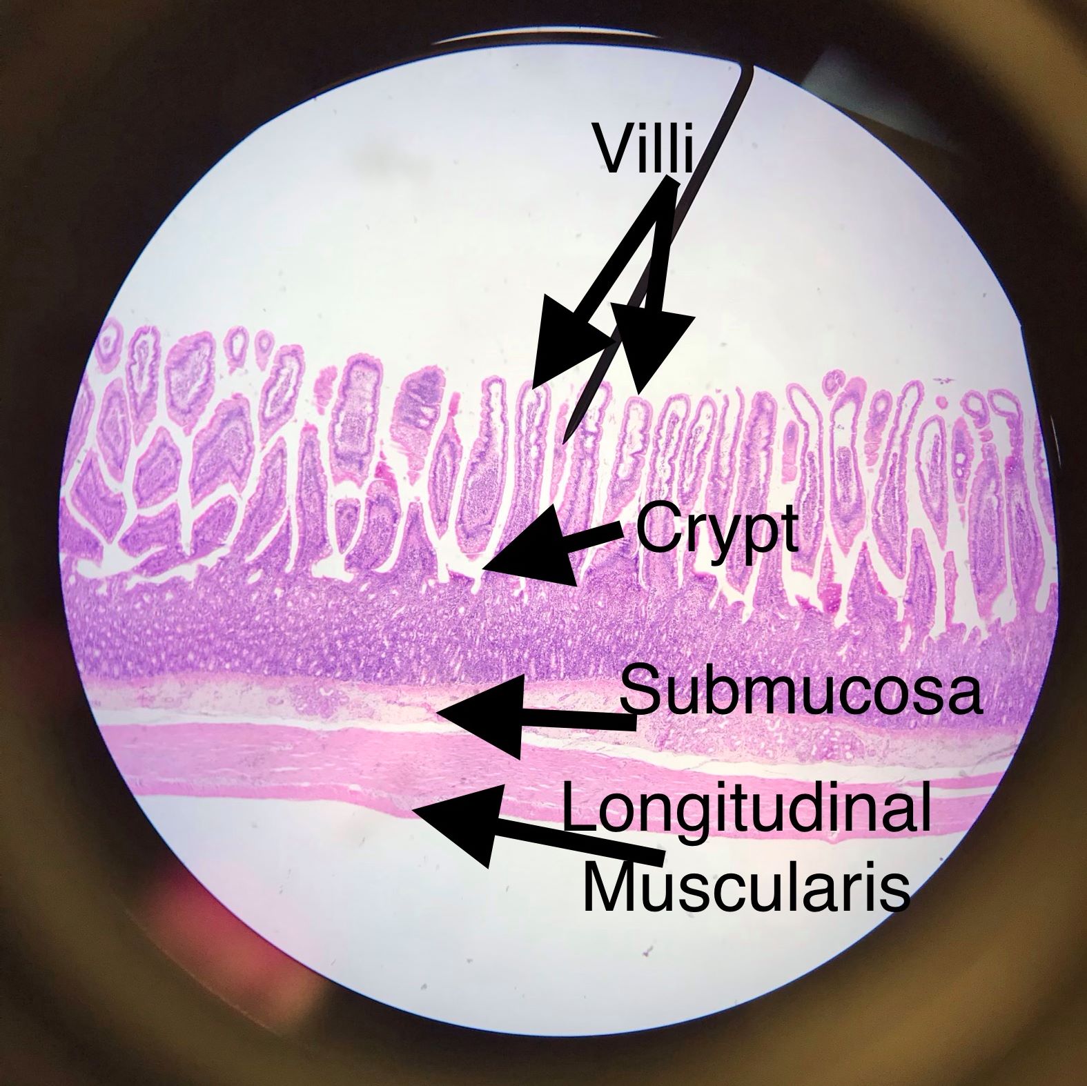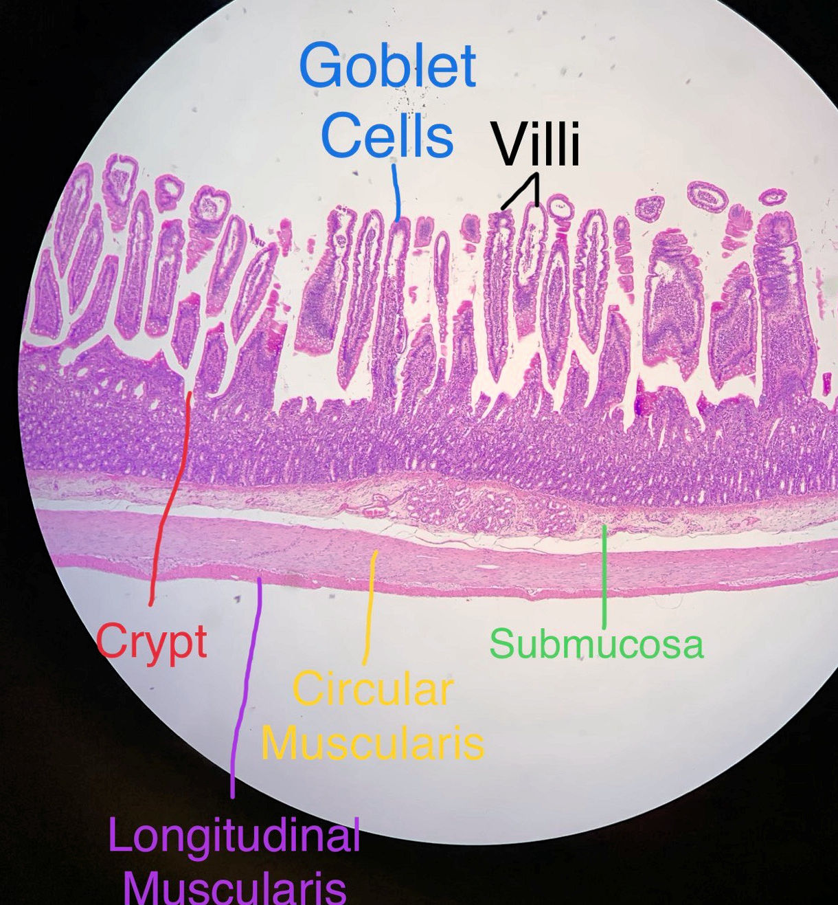
The jejunum, like the rest of the small intestine, has mucosal, submucosal, muscularis, and serosal layers. Furthermore, the muscularis is divided into an inner circular and outer longitudinal layer. The Jejunum has huge villi with crypts at their base. The crypts contain stem cells that replace cells lost through normal wear and tear. There is not much in the submucosal layers since it lacks both duodenal glands and Peyer's patches. Be sure to look at this under high power too.
These pictures were taken by bio 139 students in the spring of 2018 and fall of 2019. Scroll through the pictures and compare them with the labeled picture. Select one and draw it. This model shows what the layers should be also. It is sort of a combnation of all 3 sections though.
| Lab Book Image | Student Images |
|---|---|
|
|
|