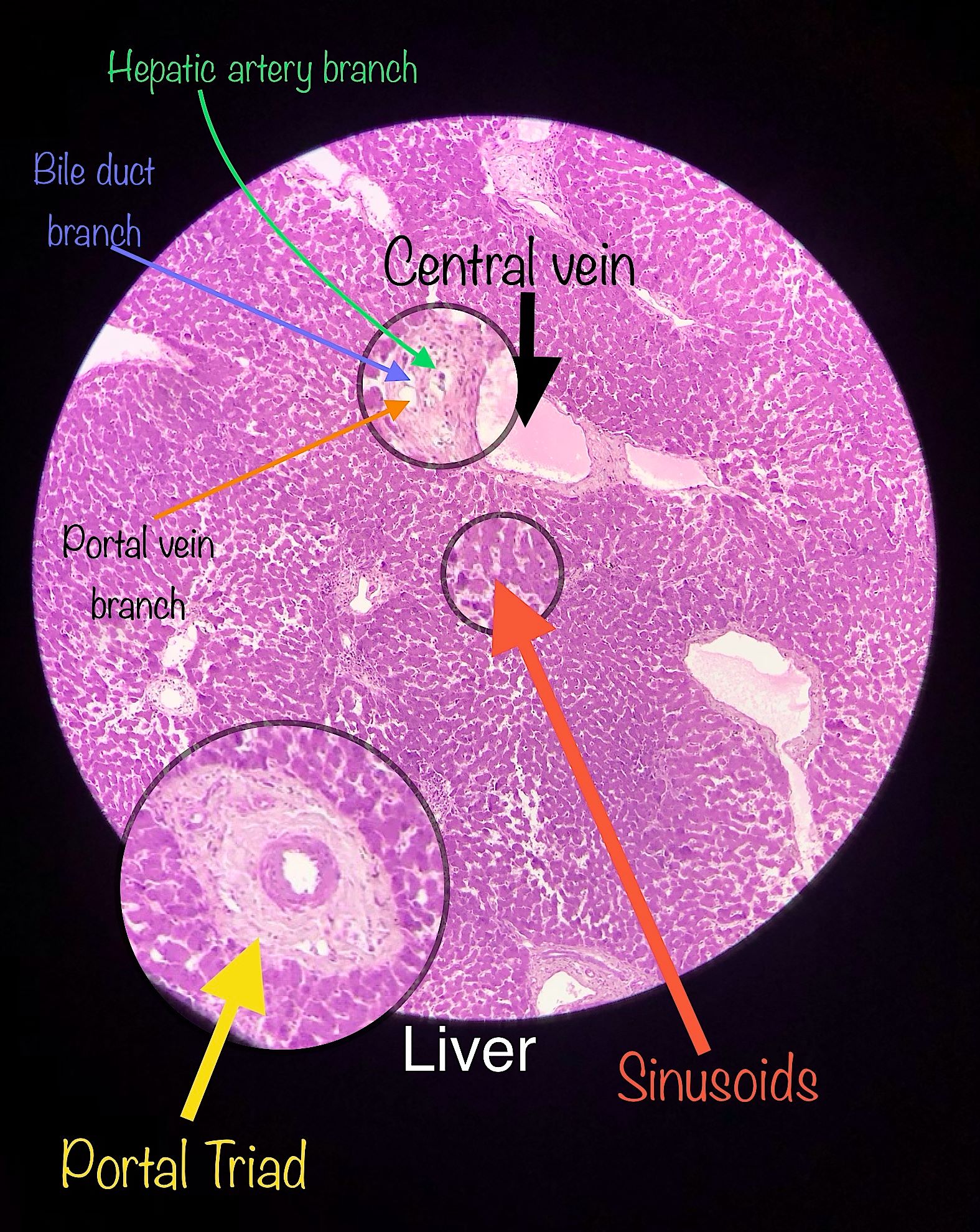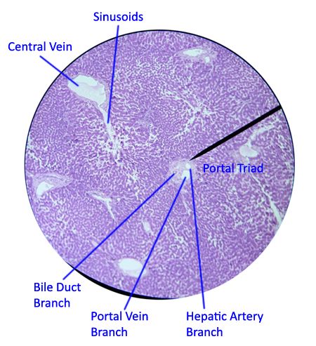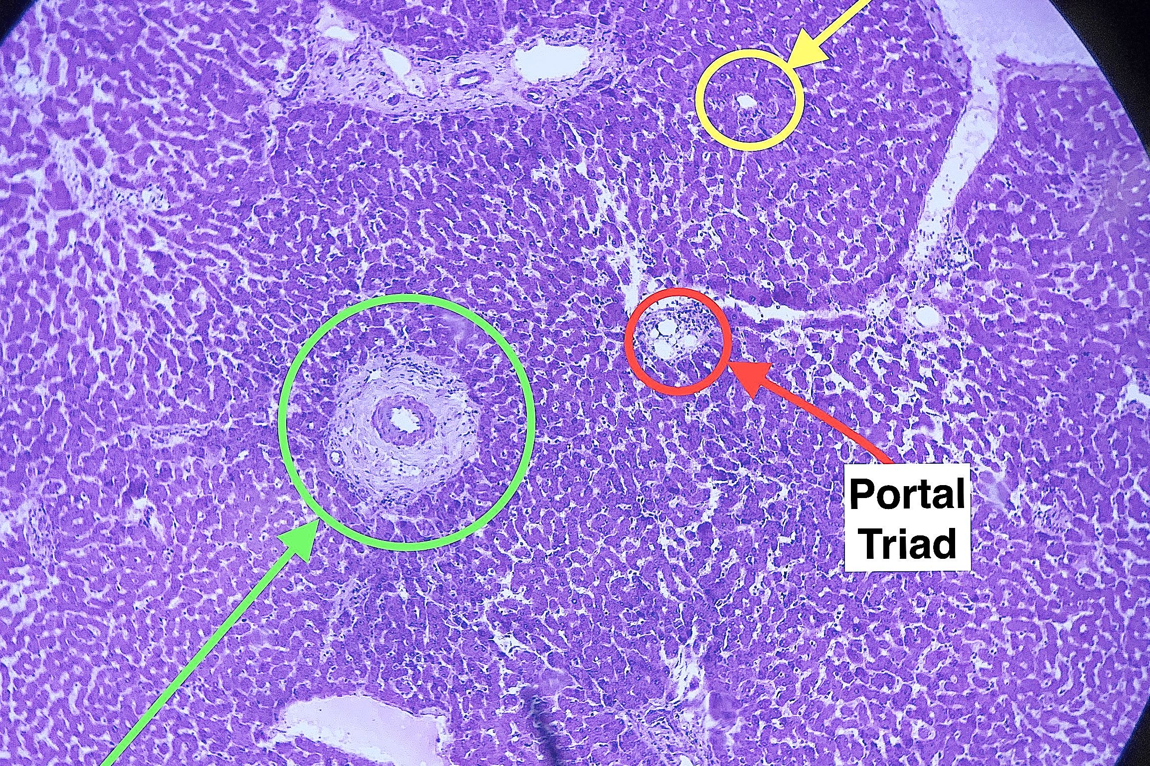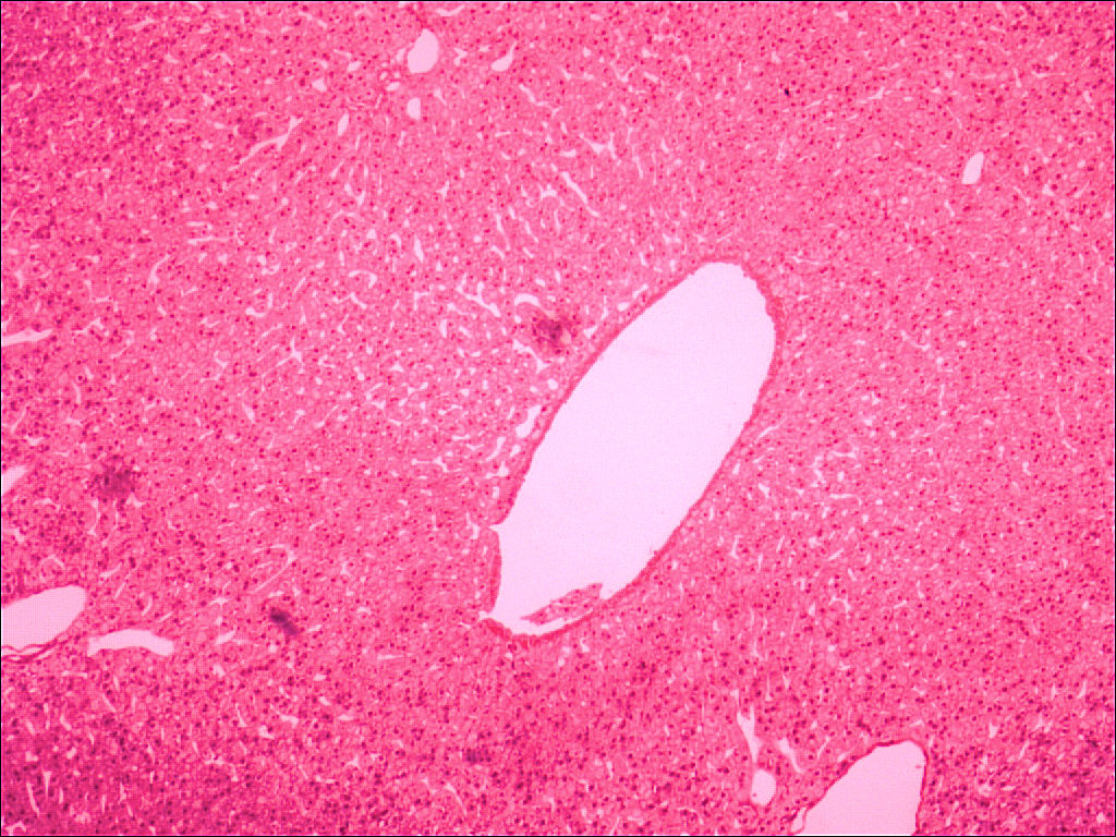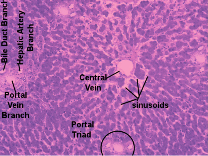
The cells of the liver are encapsulated into hexagonal shaped lobules. At each corner is a portal triad while in the center is the central vein. The portal triad is made up of 3 small vessels. An arteriole from the hepatic artery and a venule from the portal vein bring blood to the lobule. The blood drains through a series sinusoids that are lined by hepatocytes. The hepatocytes absorb substances from the blood from the sinusoids and process it; either to be secreted from the body, stored in the liver, or repackaged and secreted into the sinusoid. Substances secreted into the sinusoid along with the blood drain into the central vein of the lobule which will eventually become part of the hepatic vein. Hepatocytes will secrete phospholipids, bile acids, and bile pigments into other structures called bile canaliculi. These canaliculi lead to the bile duct and eventually exit through the hepatic duct where they are stored in the gall bladder. The phospholipids and bile acids help to digest lipids while the bile pigments are waste products (such as bilirubin) that the liver is getting rid of. Lining the sinusoids are specially macrophages called Kupffer cells. These cells recycle red blood cells and capture the iron at the center of the hemoglobin.
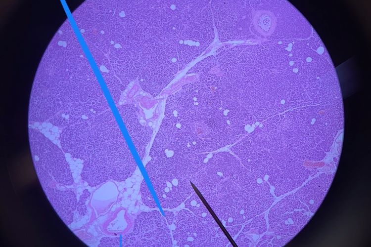
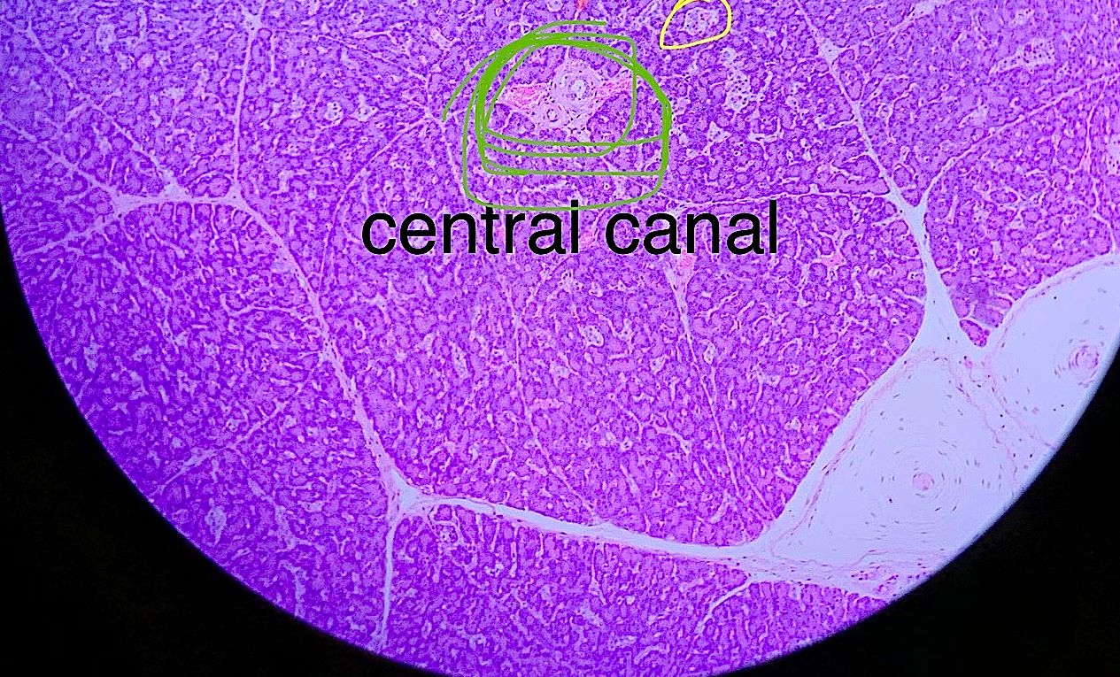
These pictures were taken by bio 139 students in the spring of 2018 and fall of 2019. Scroll through the pictures and compare them with the labeled picture. Select one and draw it. This picture is a model of what you are looking at.
| Lab Book Image | Student Images |
|---|---|
|
|
|