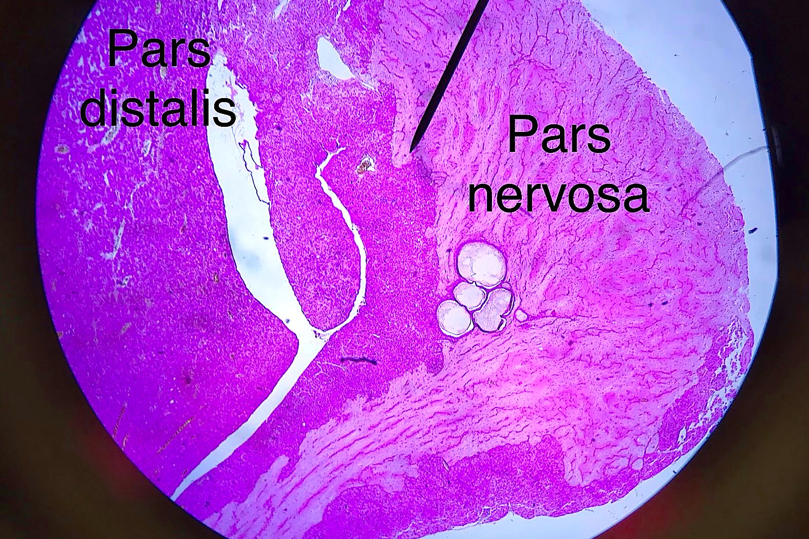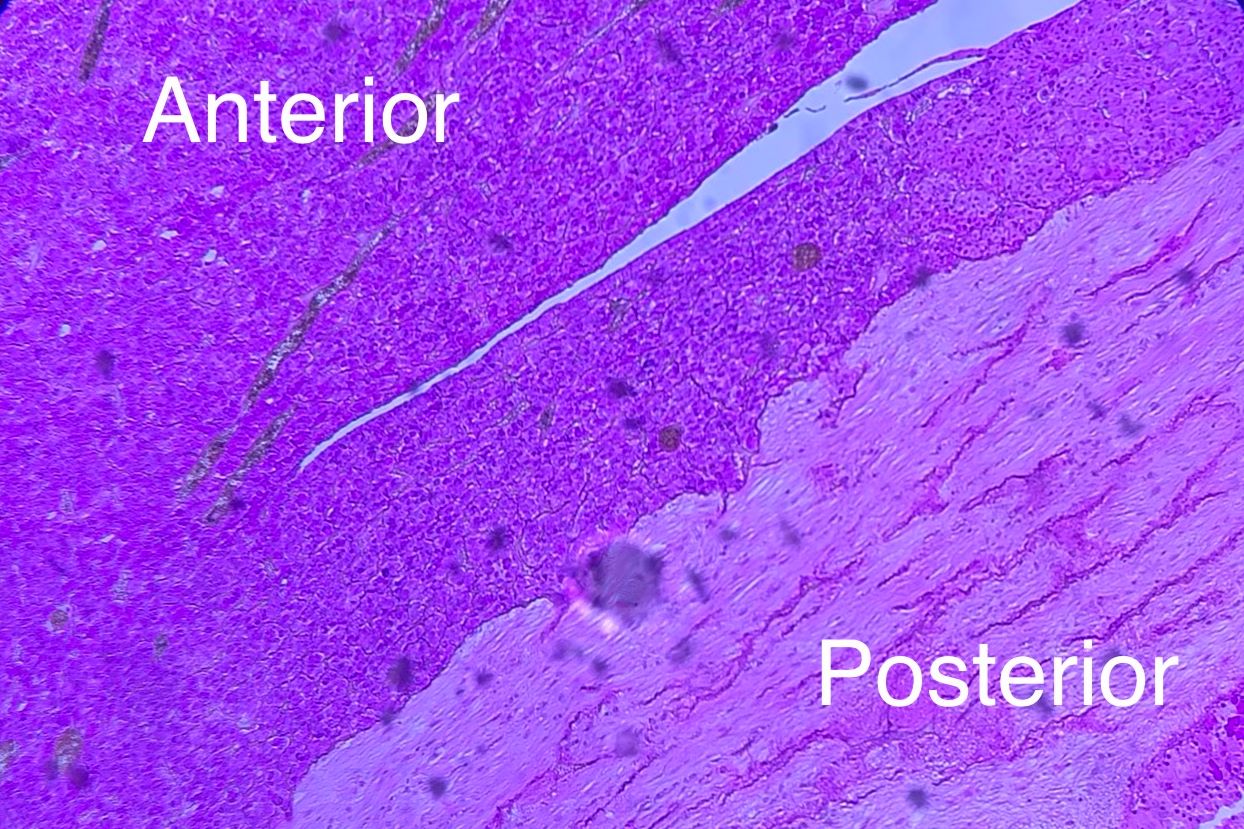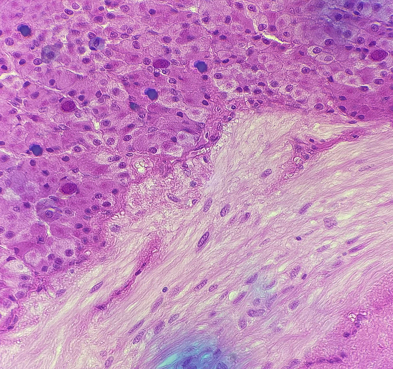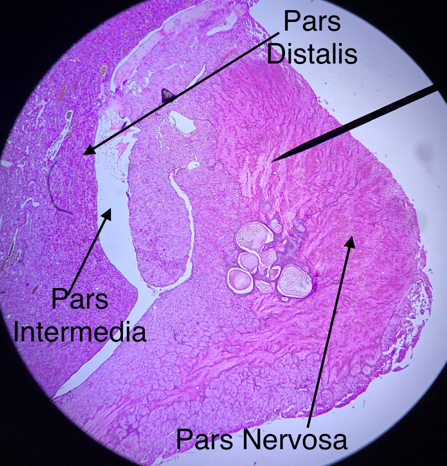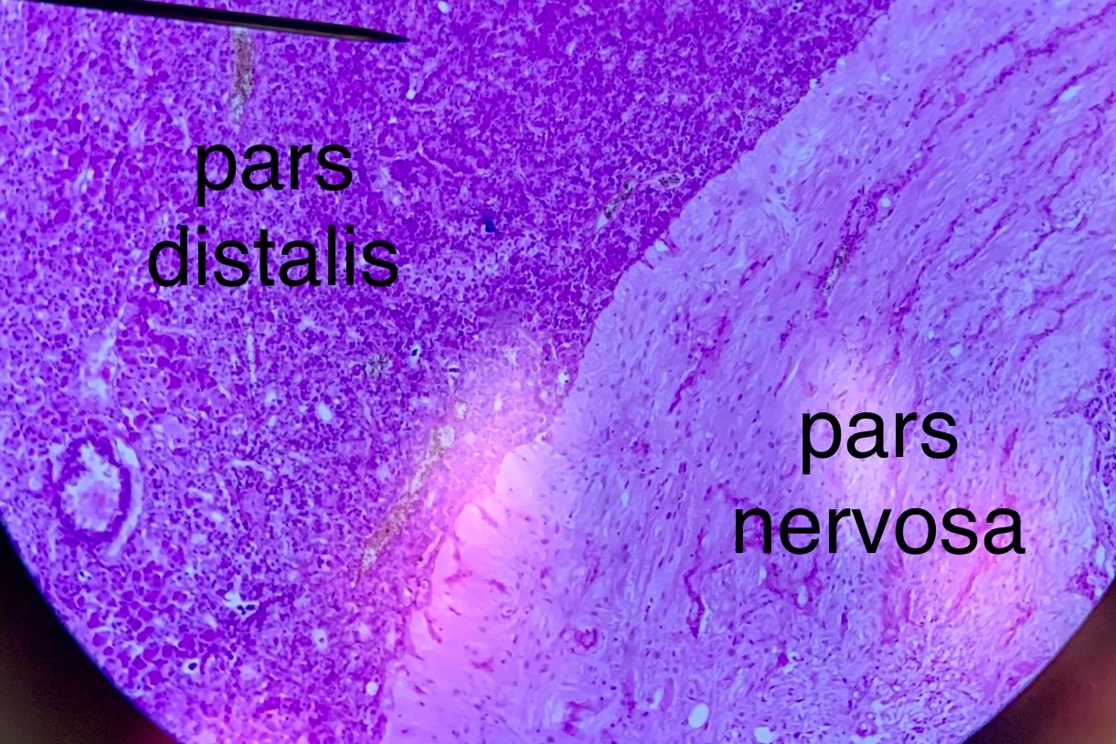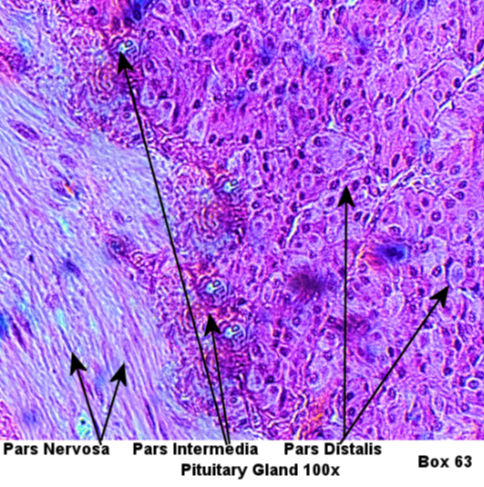
This page has endocrine histology of the pituitary gland under low power. The pituitary gland is attached to the hypothalamus via a structure called the pituitary stalk or infundibulum which consists of axons leading from the hypothalamus to the posterior pituitary gland. Therefore, the posterior pituitary gland mostly resembles white matter under the microscope and is is therefore called the pars nervosa due to its resemblance of nervous tissue. These axons release oxytocin and antidiuretic hormone. The anterior pituitary mostly resembles normal glandular tissue and is this region is called the pars distalis because it is further from the brain (point of attachment). A zone called pars intermedia is the transition between the pars nervosa. and pars distalis. This zone makes melanocyte stimulating hormone.
Slides which were taken by bio 139 students from spring of 2018 to fall of 2019 are in the right hand side and your lab book's picture is on the left hand side. Compare those pictures to the lab book pictures by scrolling trough the student pictures using the black arrows. Then draw the histology as instructed by your teacher.
| Lab Book Image | Student Images |
|---|---|
|
|
Pituitary Low Power |