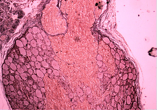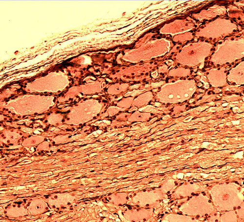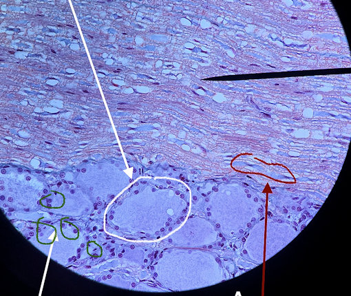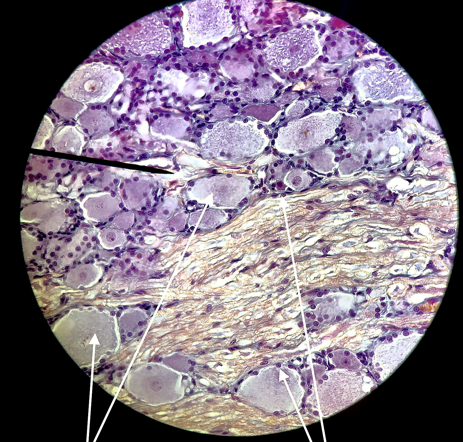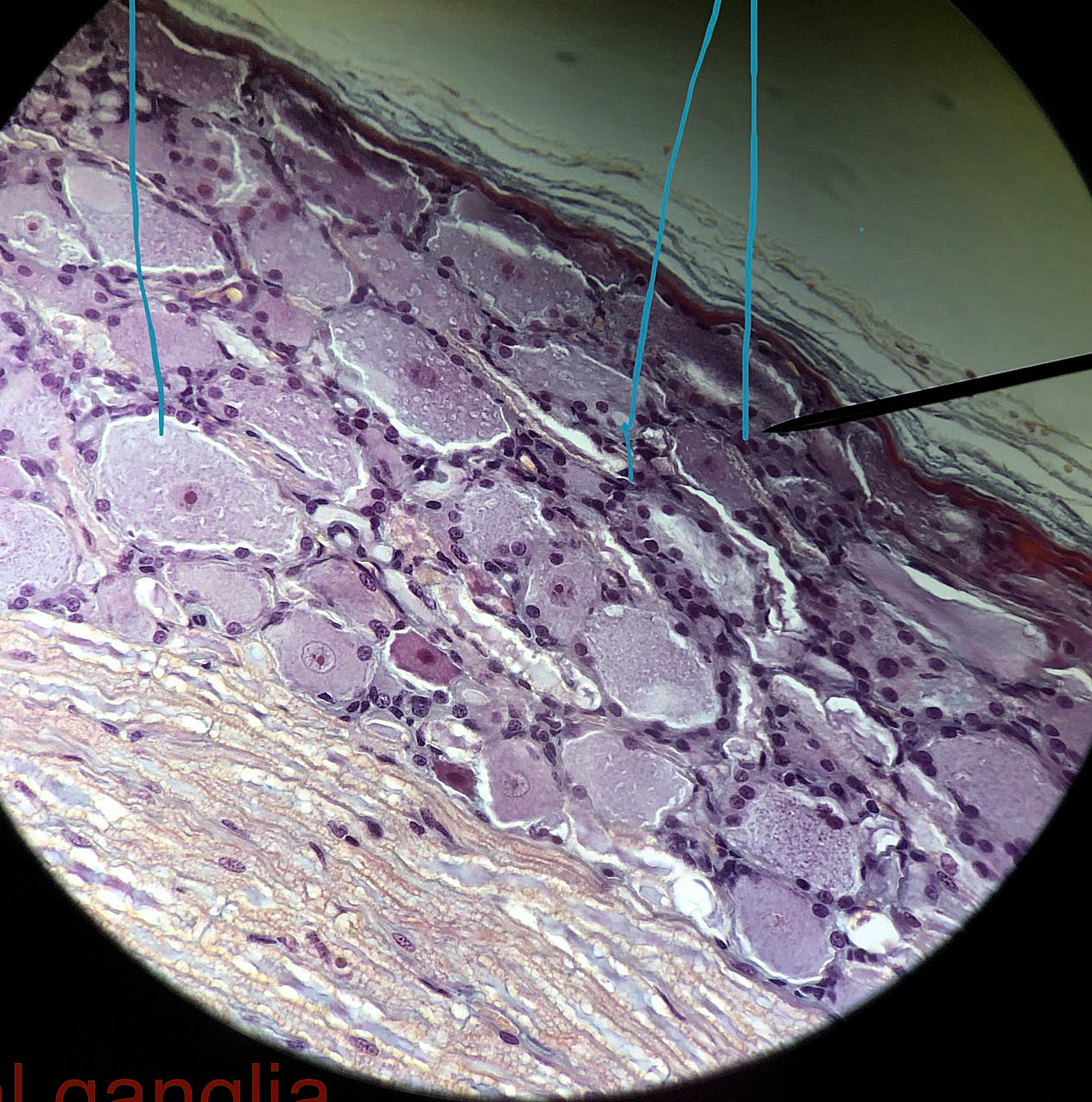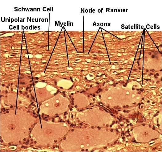
This page has histolgoy for the posterior (dorsal) root ganglia which shows unipolar neuron cell bodies and satalite cells. The picture to the left is from the lab book. The picture on the right are student files from the fall of 2019.
Nerves split when they enter the spine. Motor signals are leaving from the anterior (ventral) part (root) and sensory signals go in the posterior (dorsal) part (root). The posterior root will have a swelling that contains the cell bodies of unipolar neurons. If you were to look at the posterior horn under the microscope, you would see large unipolar cell bodies surrounded by flattened satellite cells. Satellite cells are glial cells of the peripheral nervous system that are similar to astrocytes in that they nourish and protect cell bodies in ganglia. They also form a sheath around the cell bodies that isolate them from the rest of the body. On high power, you can observe the Schwann cells. Schwann cells wrap round axons like an onion. They then produce a glycolipid called myelin which insulates the neurons. This prevents ions (sodium, potassium, calcium, and chloride) from reaching the cell membrane and altering the electrical potential of the neuron. As a result, electrical signals that are carried by the axon jump from the gaps between Schwann cells, which are called nodes of Ranvier.
Scroll through the student pictures using the arrows. Try to identify the neurons and the glial cells. Your teacher may have you either draw or screen shot and label.
| Lab Book Image | Student Images |
|---|---|
|
|