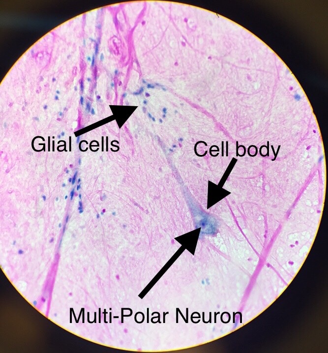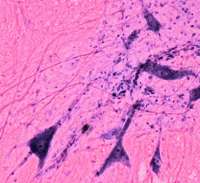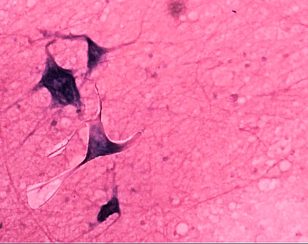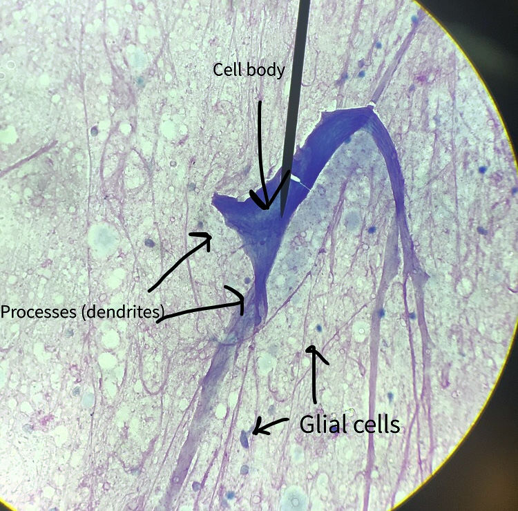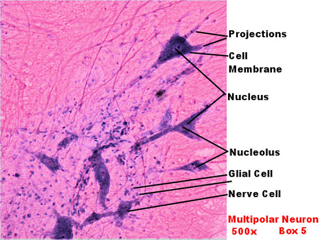
This page has Multiipoloar neurons on it. The picture to the left is from the lab book. The picture on the right are student files from the spring of 2018 to fall of 2019.
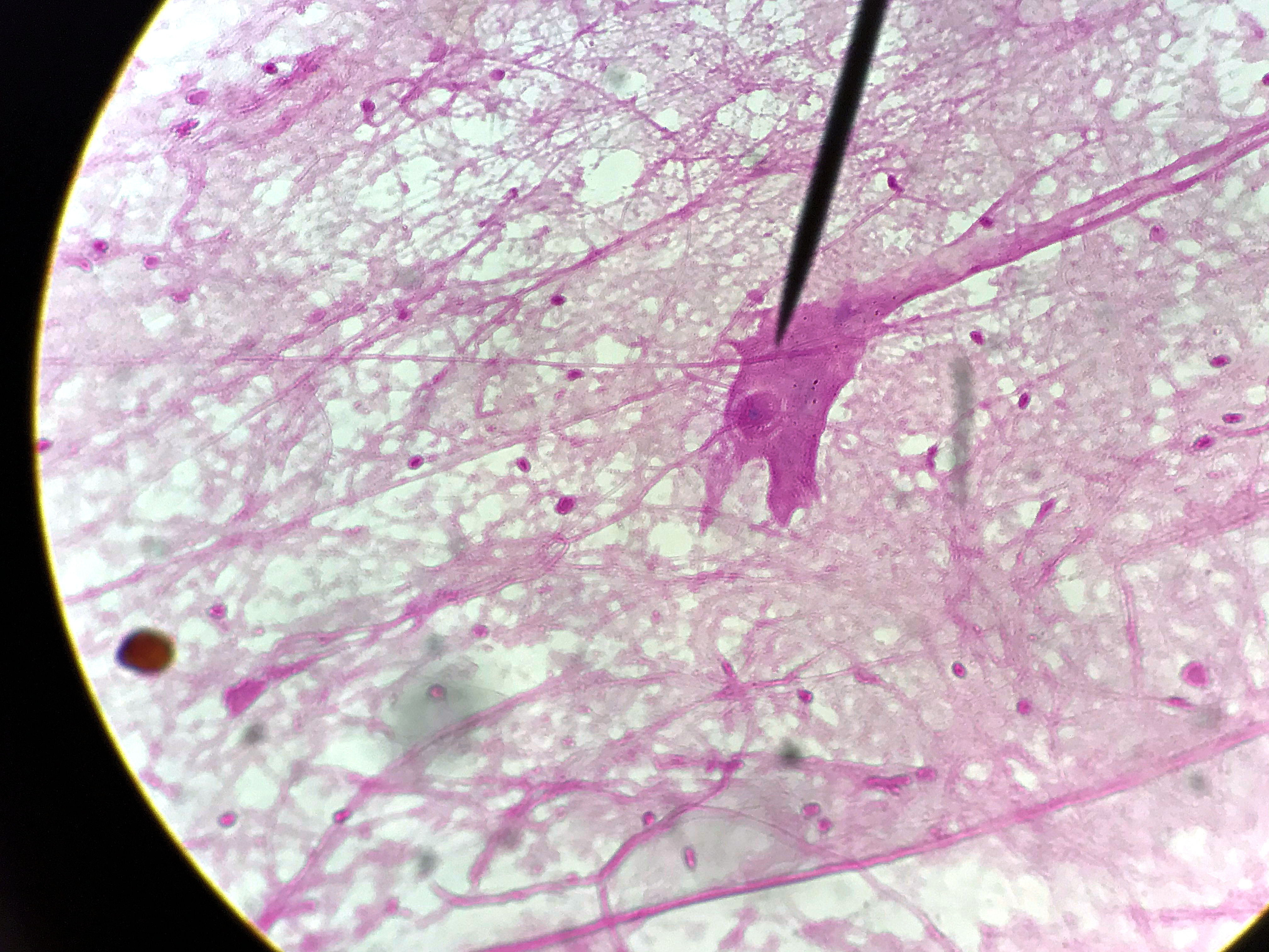
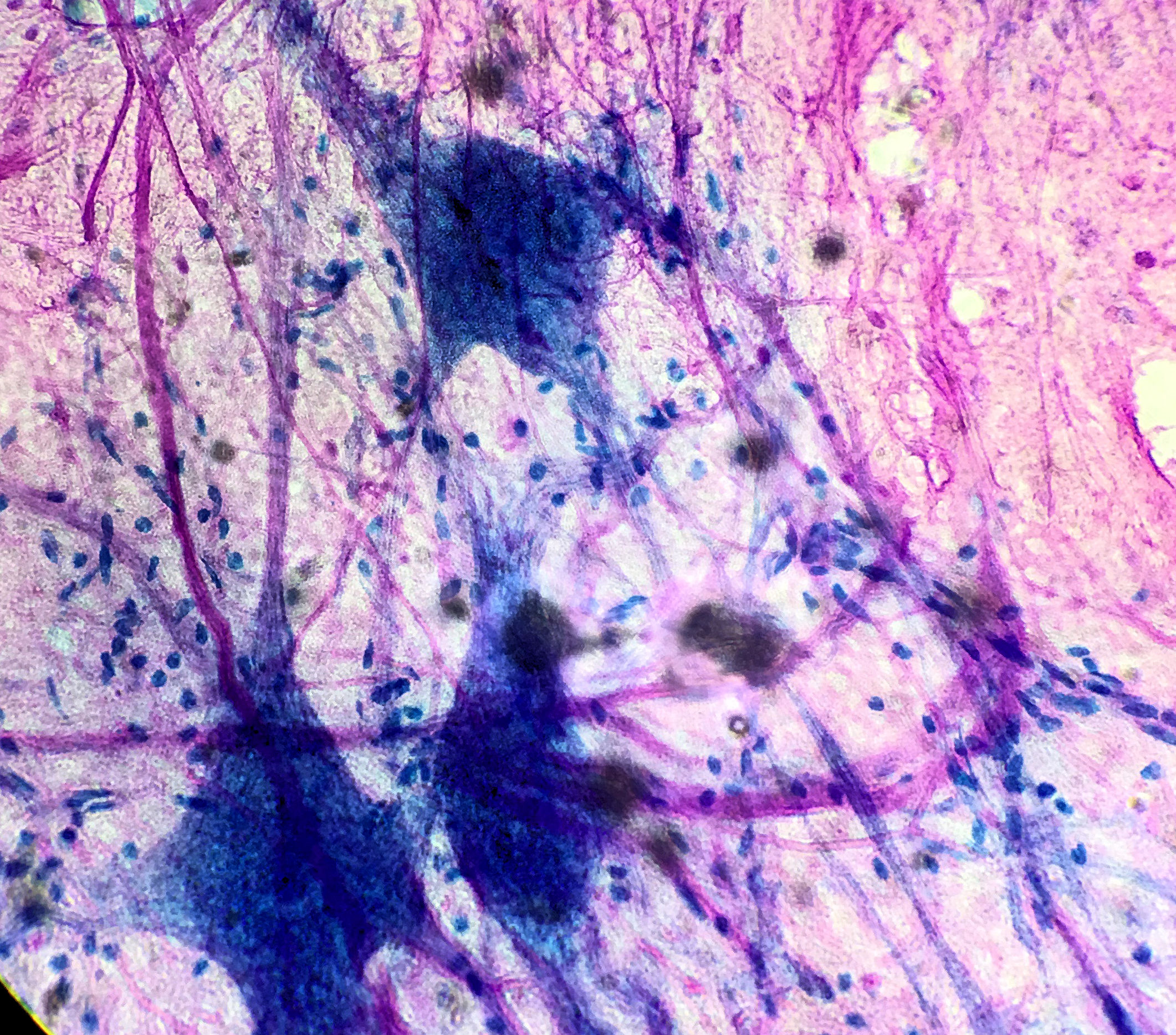
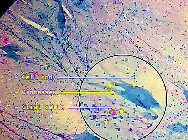
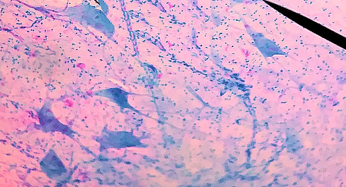
Nervous tissue is composed of excitable cells called neurons and supporting cells which are called glial cells. Neurons are typically large cells that come in many shapes. Neurons will have 1 or more processes extending from their cell body (soma) that either send or receive signals. On this page is a multipolar neuron prepared with a Nissls stain. The stain highlights the ribosomes and the nucleus. The nerve cells or neurons are the large purple cell while the supporting glial cells are the small purple dots.
Scroll through the student pictures using the arrows. Try to identify the neurons and the glial cells. Your teacher may have you either draw or screen shot and label.
| Lab Book Image | Student Images |
|---|---|
|
|