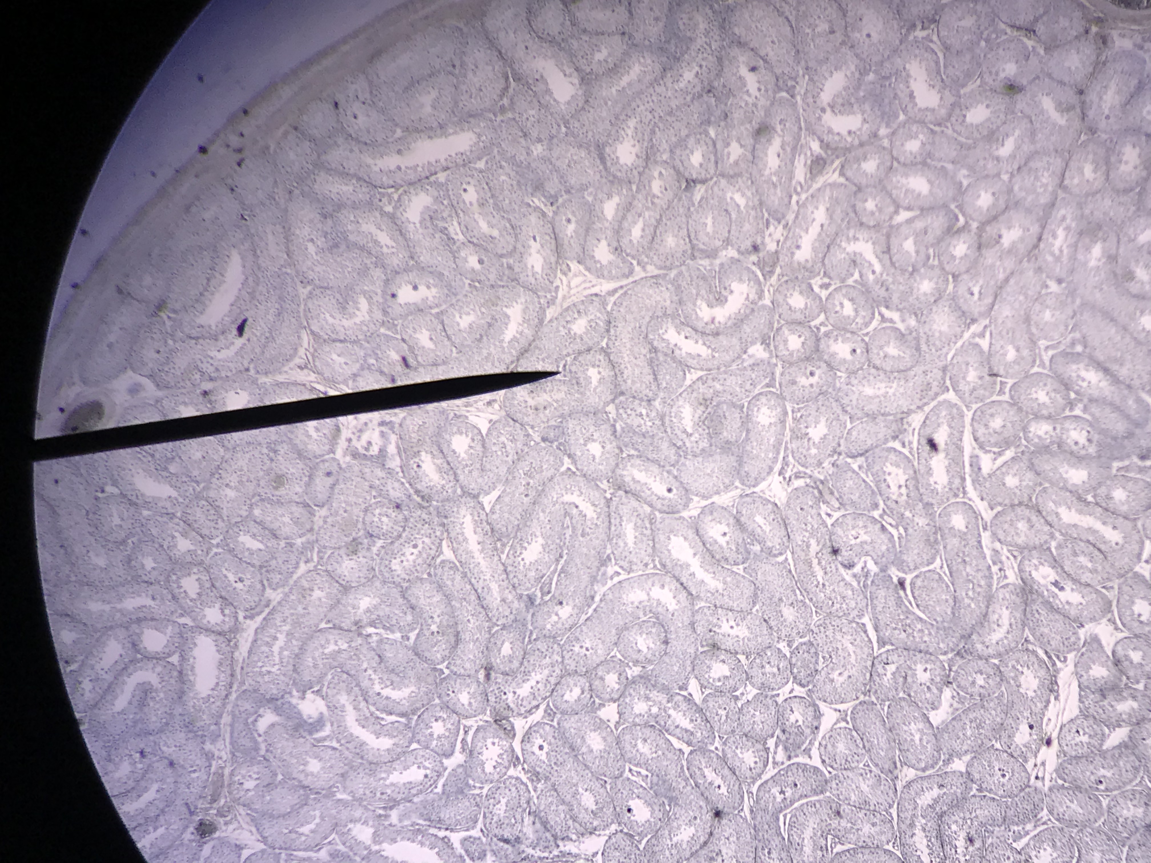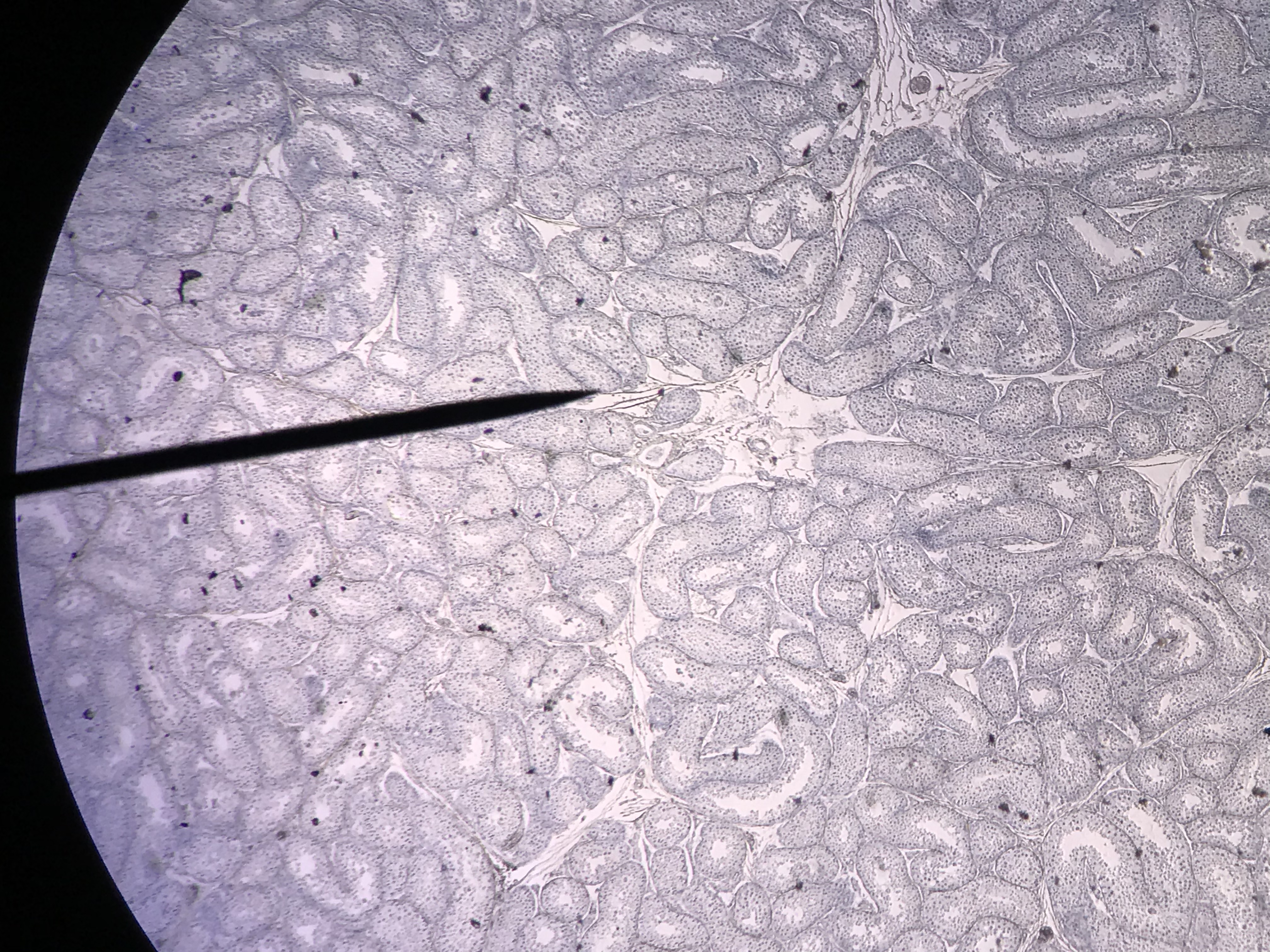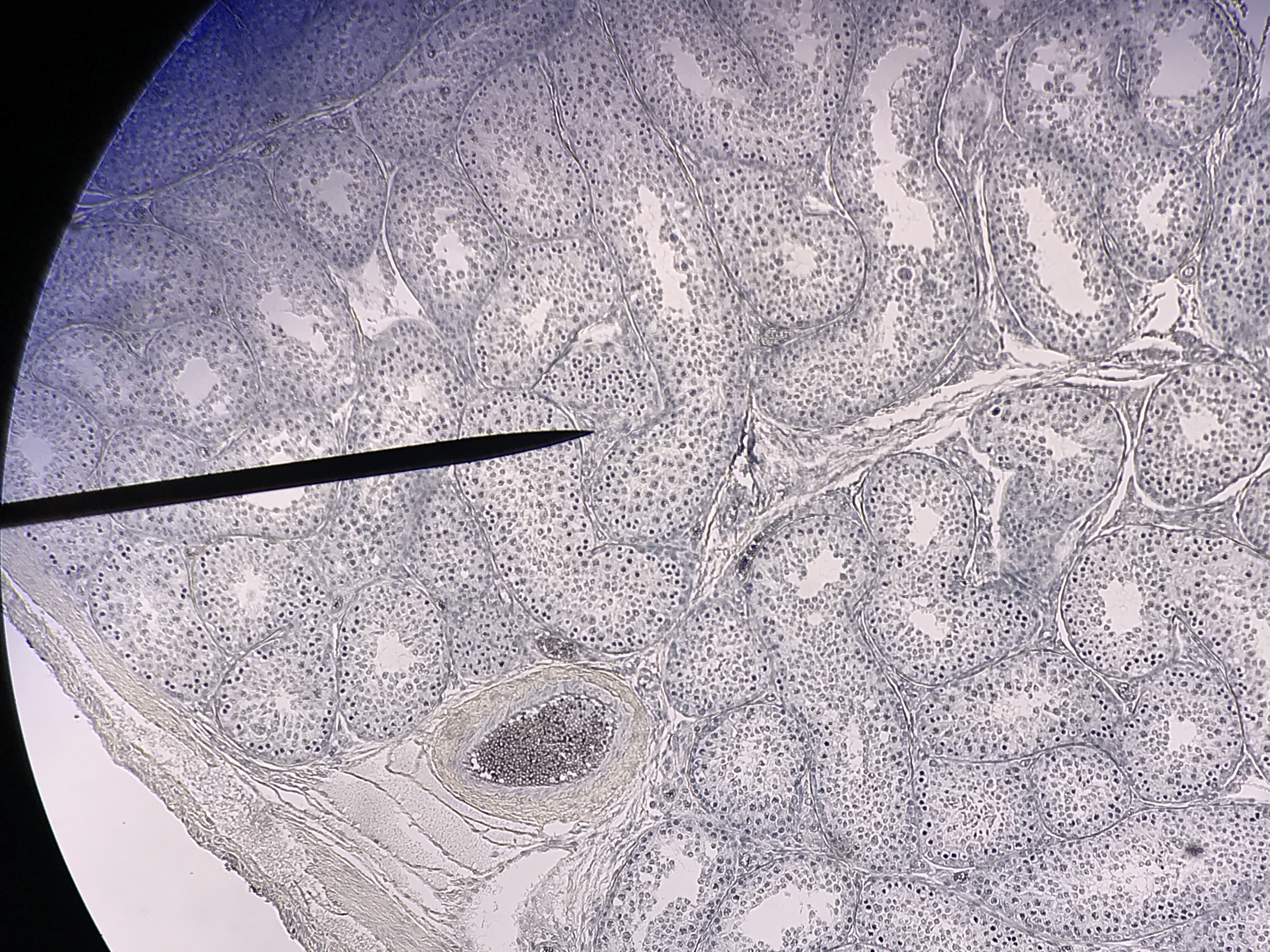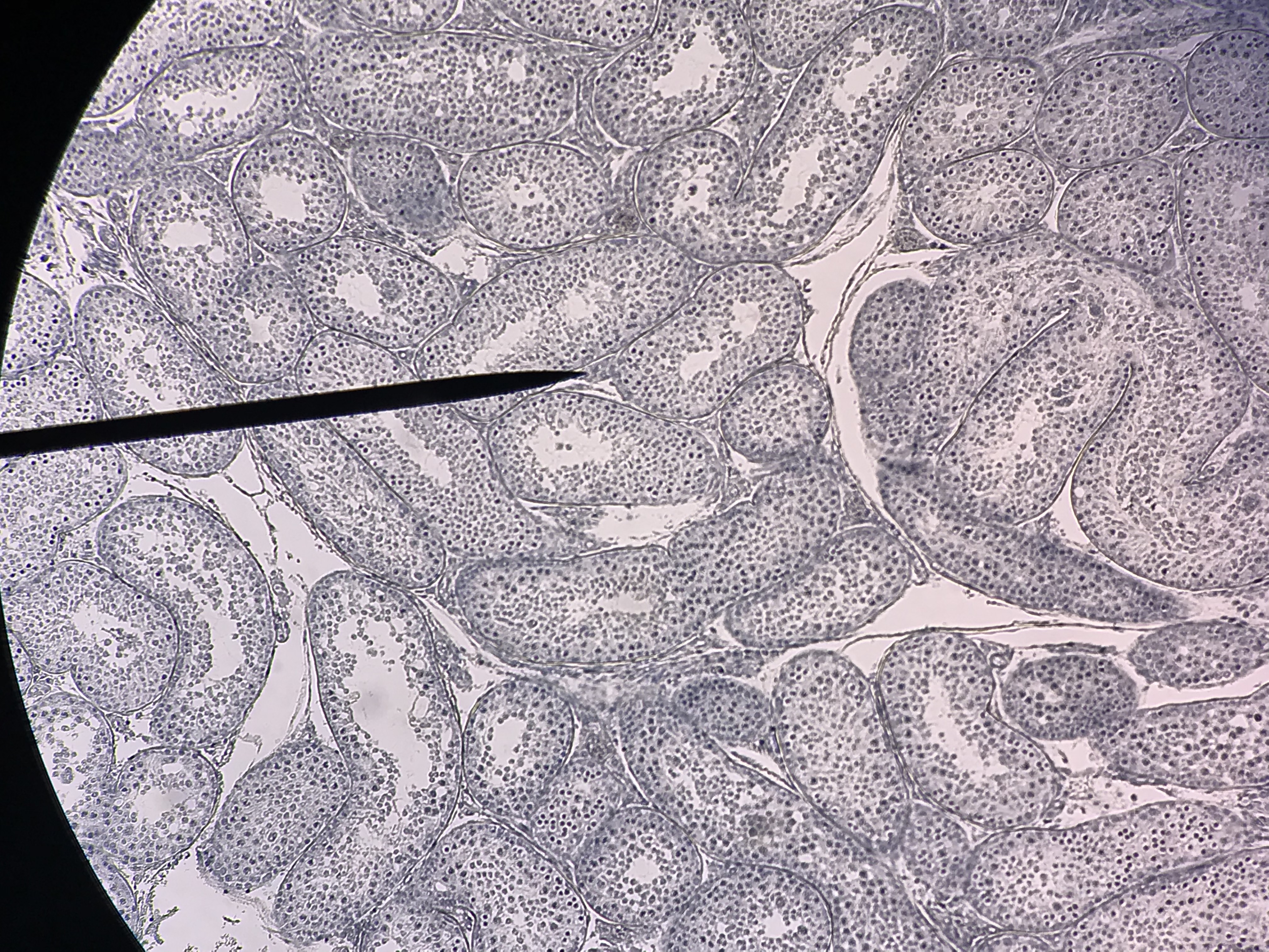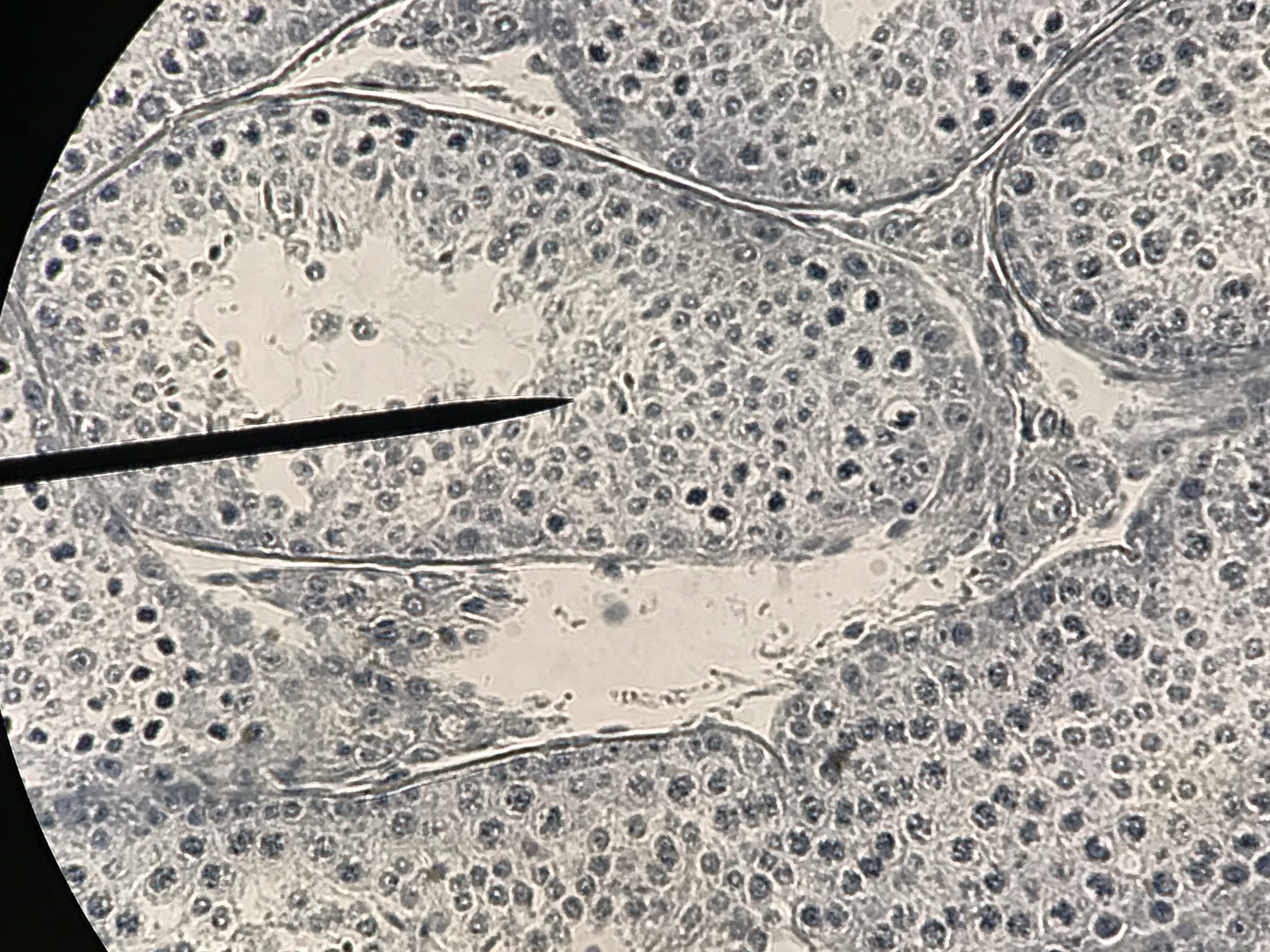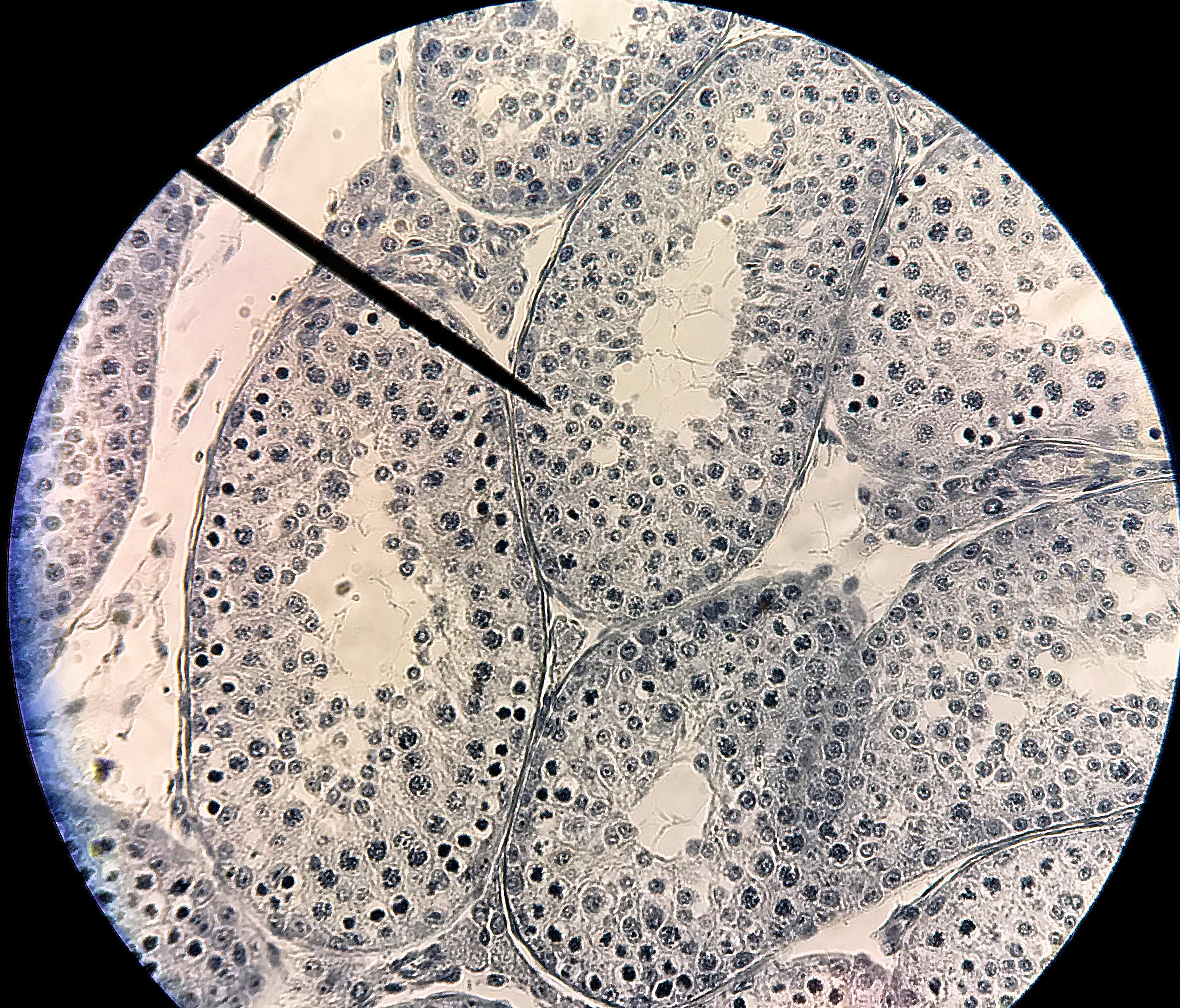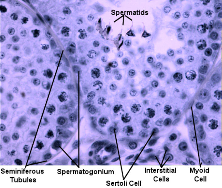
the testicles are composted hundreds of seminiferous tubules which is where spermatogenesis occurs. In a histological cross section of the seminiferous tubules, almost perfect circles can be observed. The cells on the outside of the circle are either Steroli cells that line the tubule and connect it to its basement membrane or spermatogonia. The cells in the lumen are spermatids that are destined go to the epididymis to mature into sperm. Interstitial cells are found between the seminiferous tubules. The interstitial cells function to produce testosterone which aids in gamete production, increases sex drive, and is responsible for secondary sexual characteristics such as thickening of bone, increased muscle mass and facial hair. A thin capsule actually separates the seminiferous tubules from the interstitial cells. This forms a barrier so that the man's immune system does not attach the sperm.
These pictures were taken by me in the spring of 2021. They progress from scanning power (40x) to high power (400x). Go through the pictures. Select one, draw it, and label the layers.
| Labeled Image | Unlabeled Images |
|---|---|
|
|