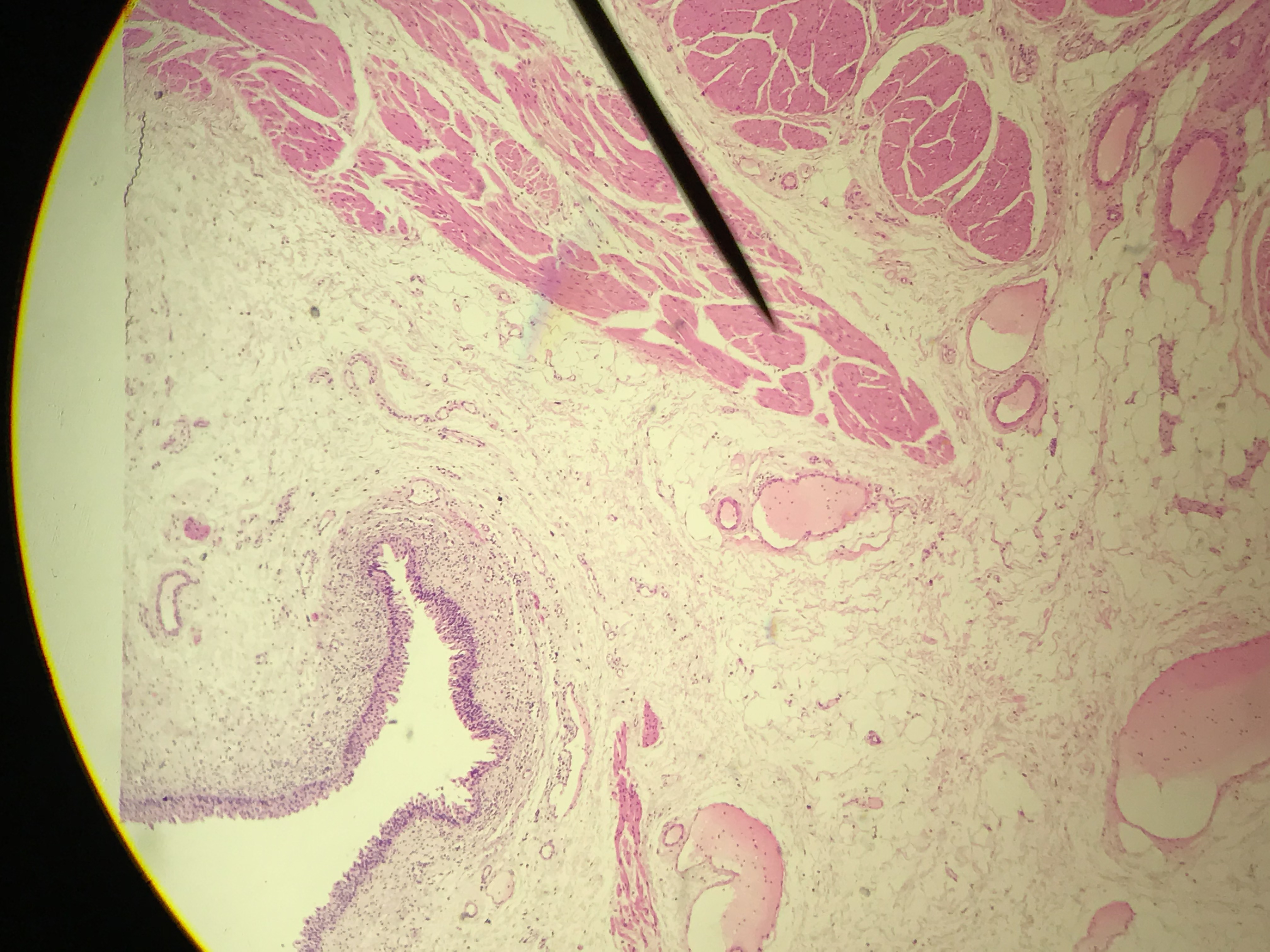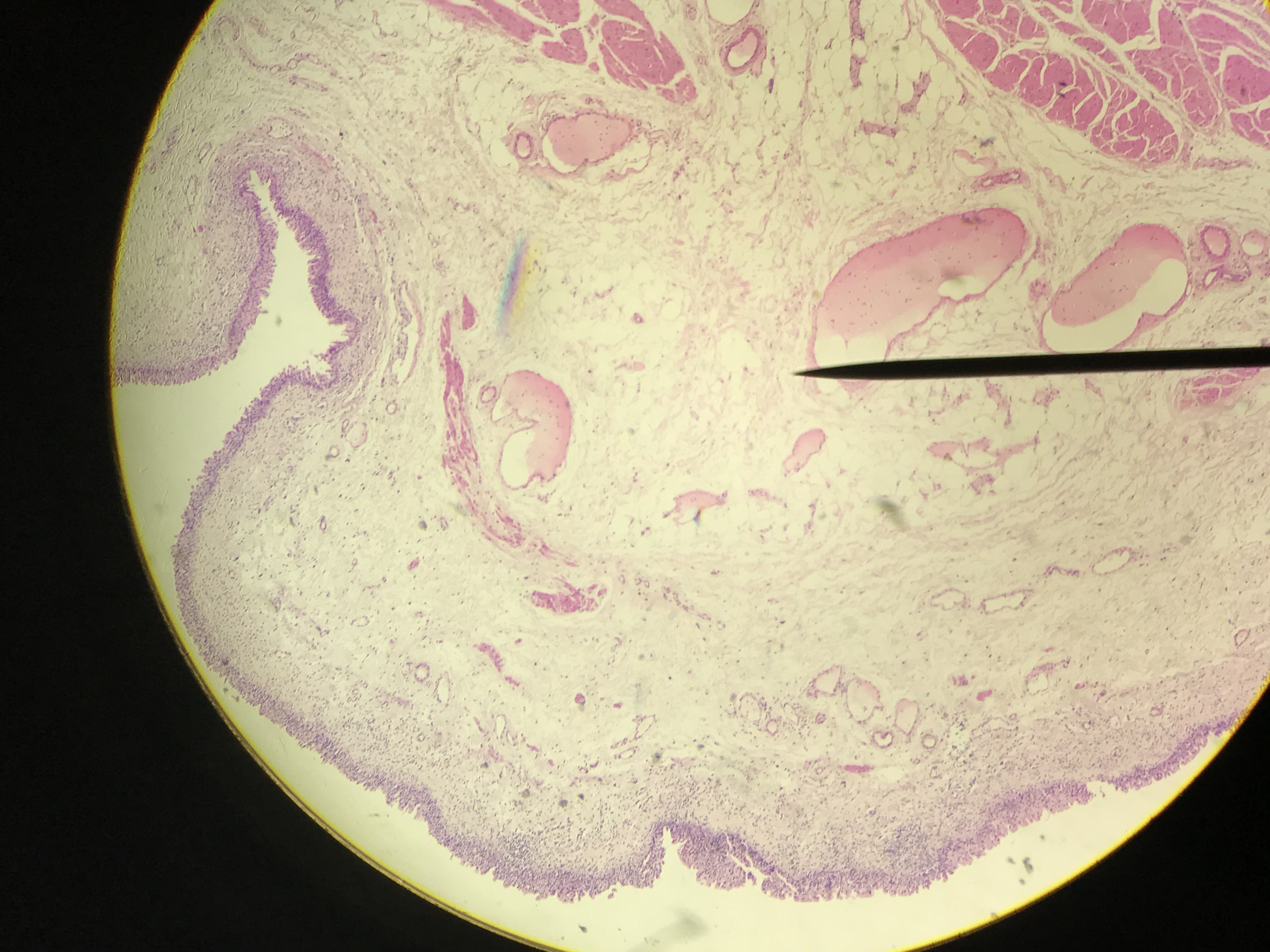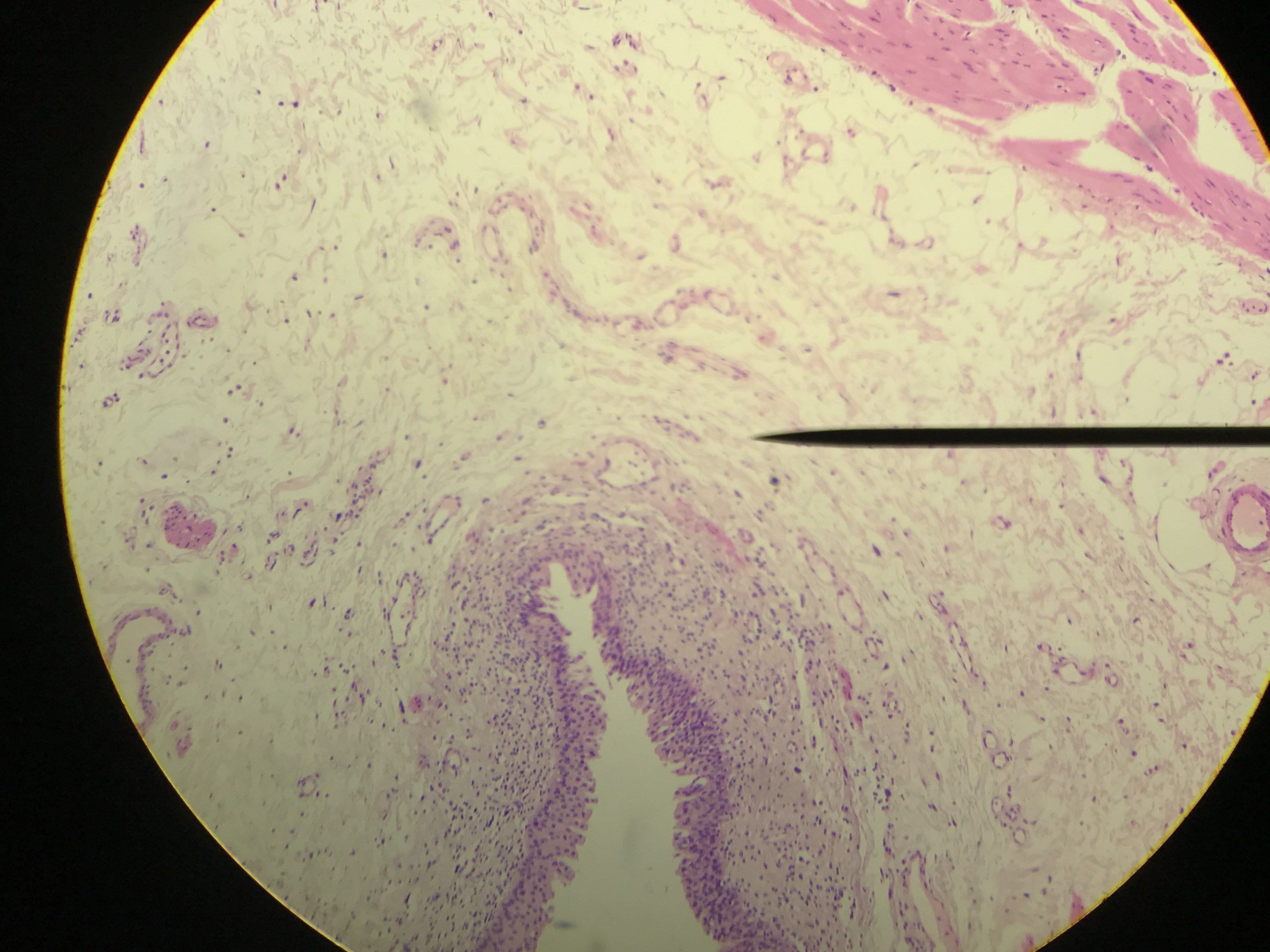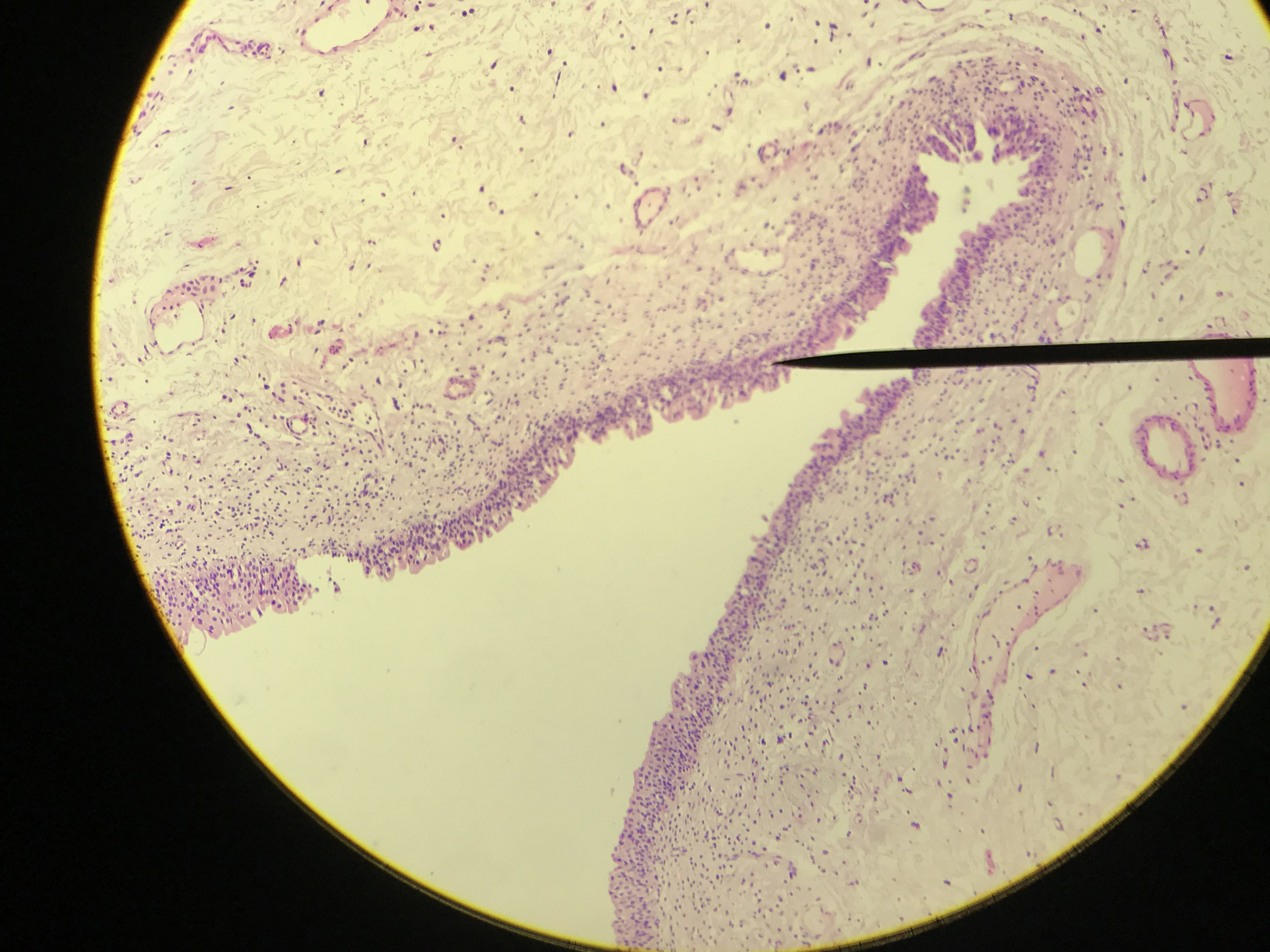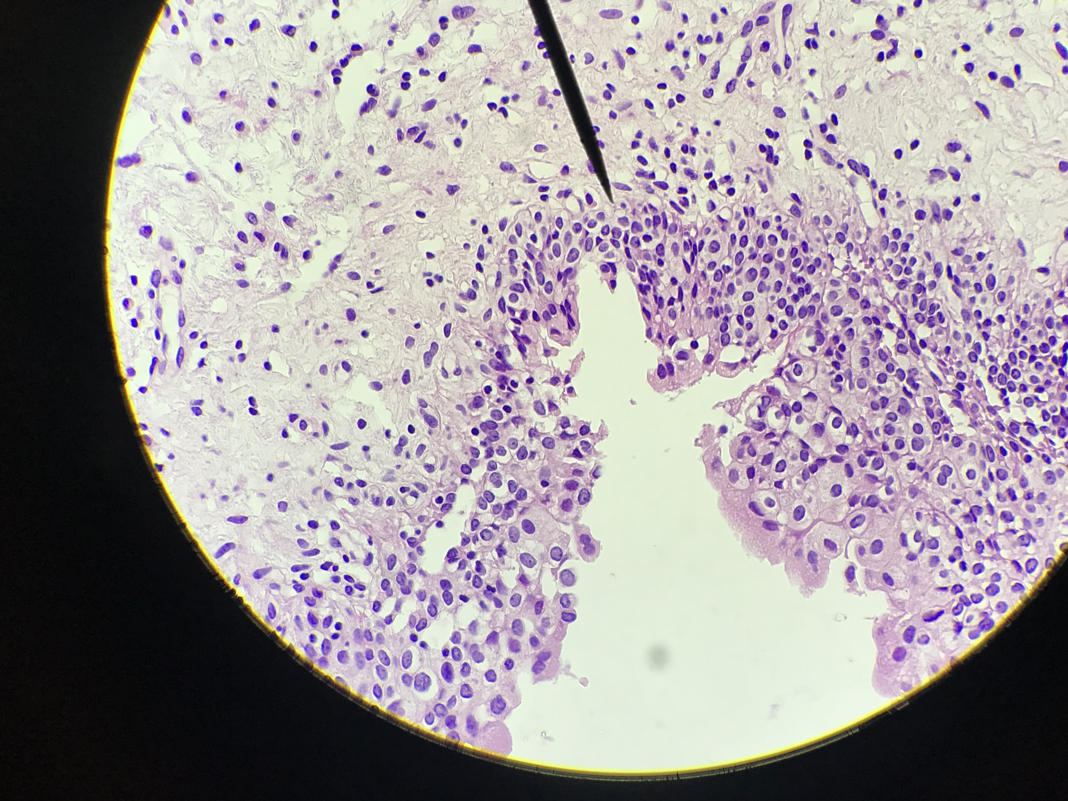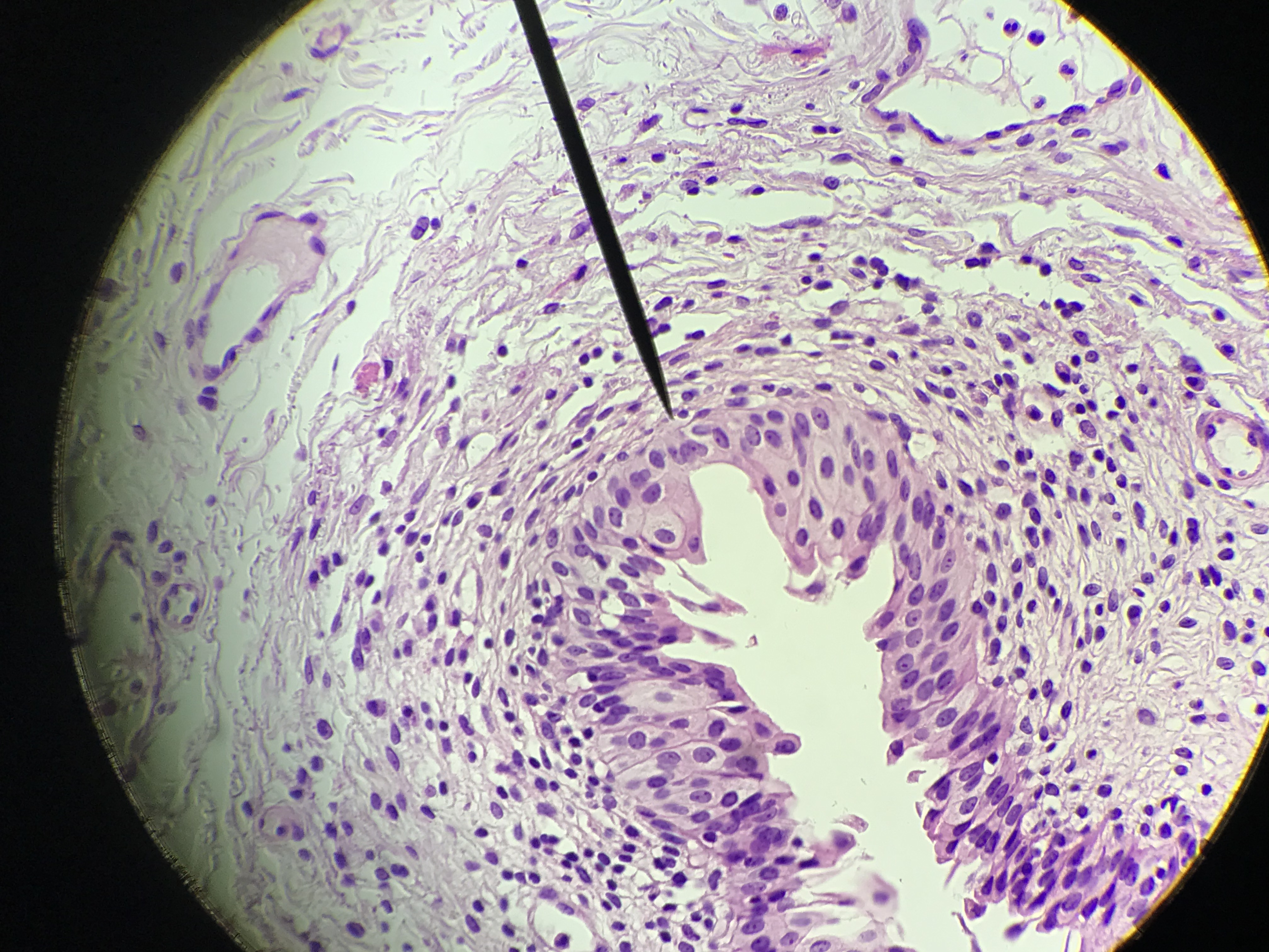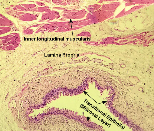
The Uirnary Bladder has 4 distinct layers of tissue which form the mucosal, lamina propria, muscularis and serosa. The Uirnary Bladder is lined with transitional epithelial tissue. This allows for distension (expansion) as urine flows through the Uirnary Bladder. The lamina propria is deep the mucosal layer and is made up of a mix of areolar and elastic connective tissue. Opposite to the digestive system, the muscularis is made up of an inner longitudinal layer then a circular layer and lastly an outter longitudinal layer. These layers propel the urine by peristaltic contractions. Lastly, a serosa layer wraps the Uirnary Bladder.
These pictures were taken by me in the spring of 2021. They progress from scanning power (40x) to high power (400x). Go through the pictures. Select one, draw it, and label the layers.
| Labeled Image | Unlabeled Images |
|---|---|
|
|