Human Anatomy Histology
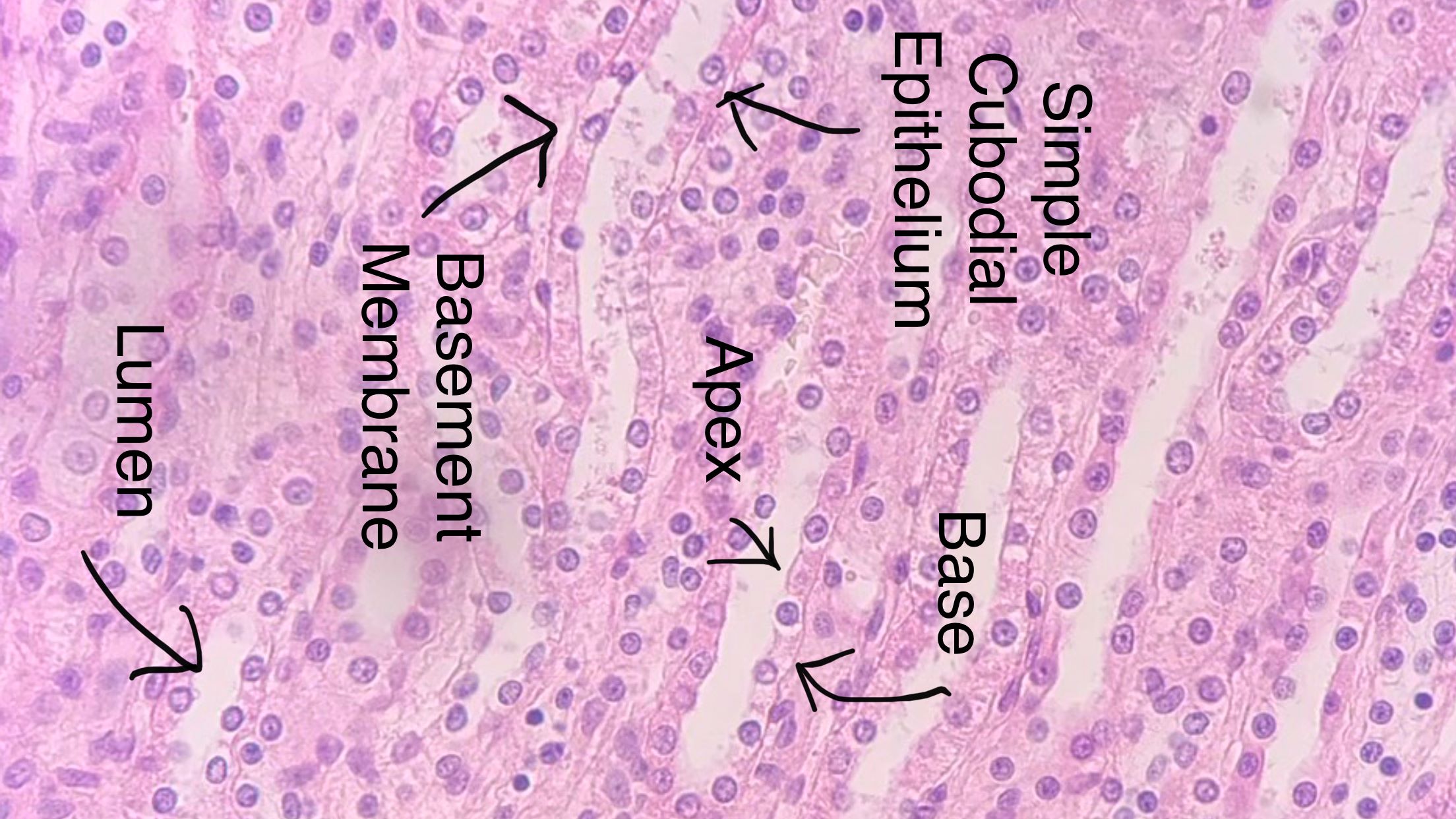
Simple Cuboidal
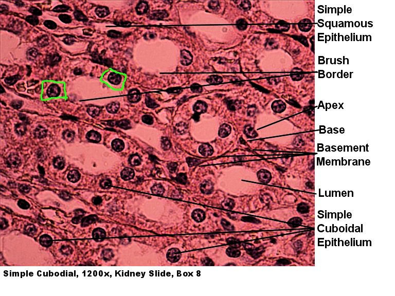
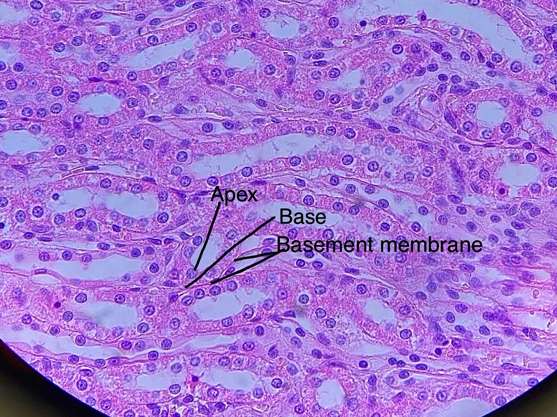
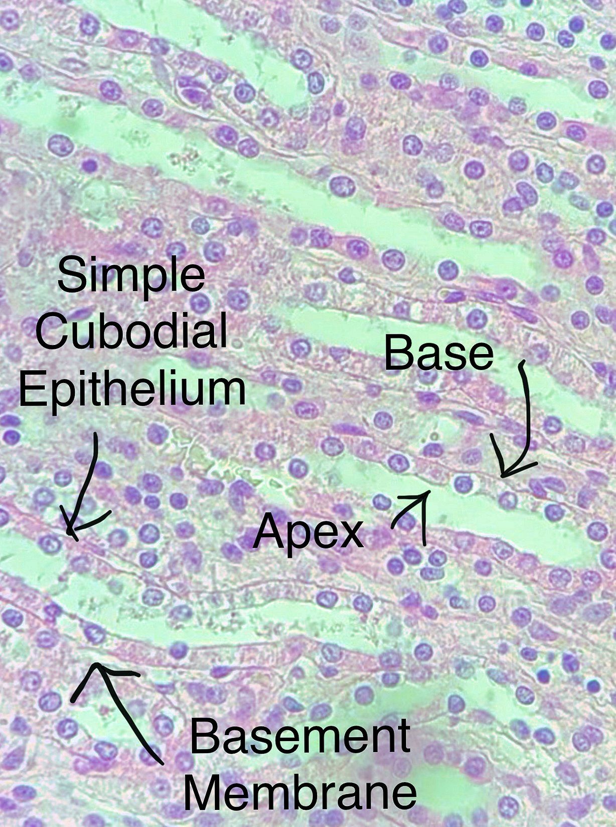
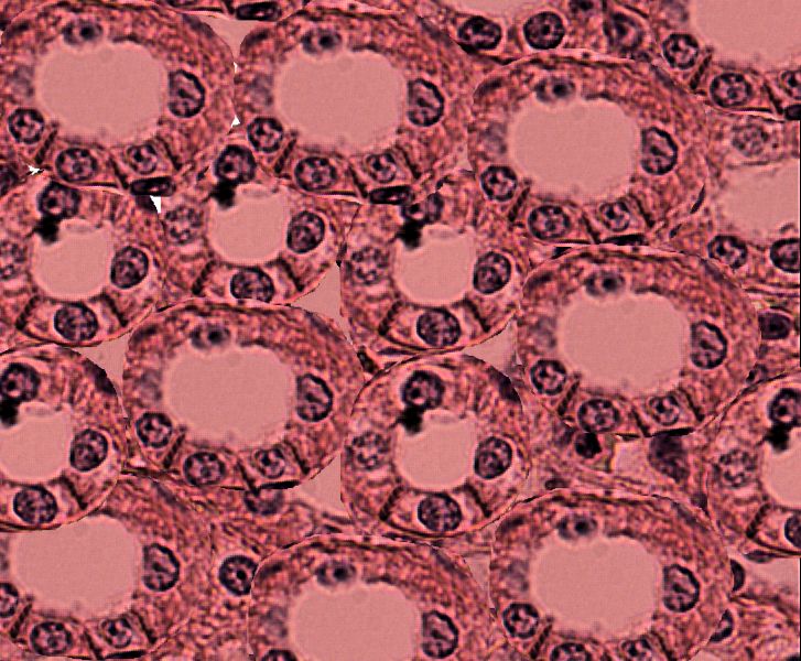
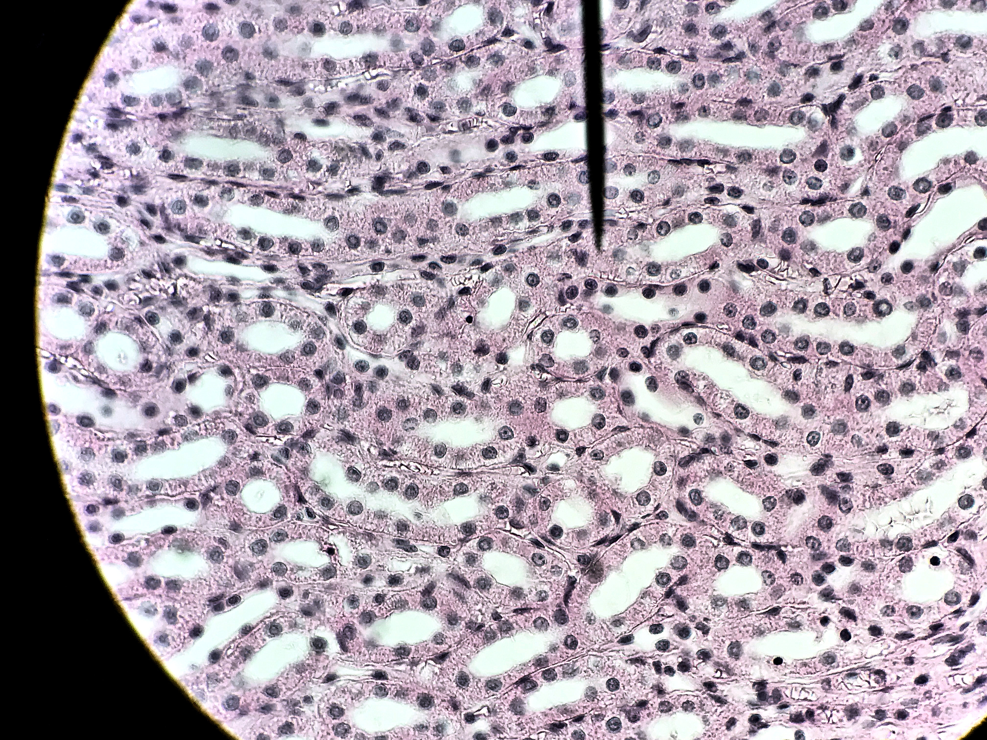
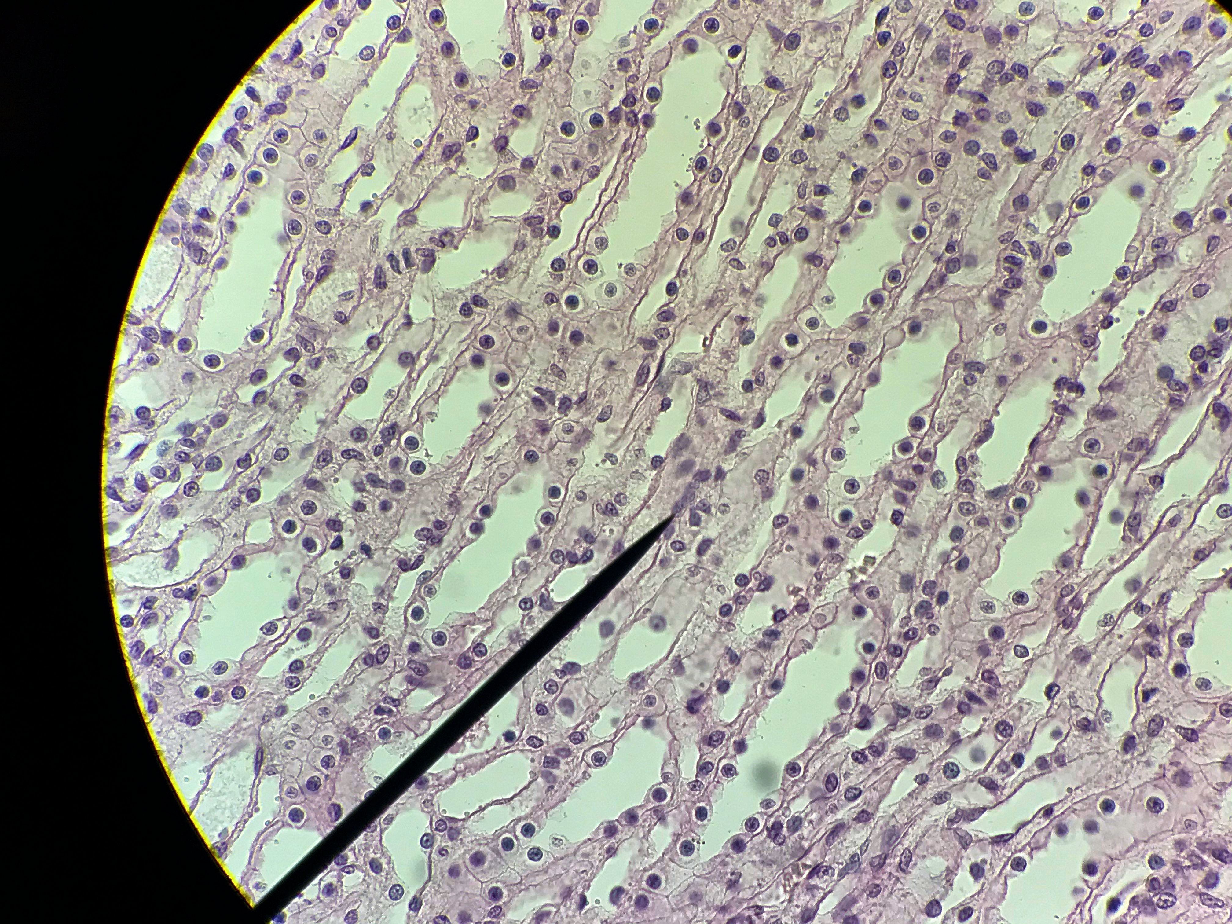
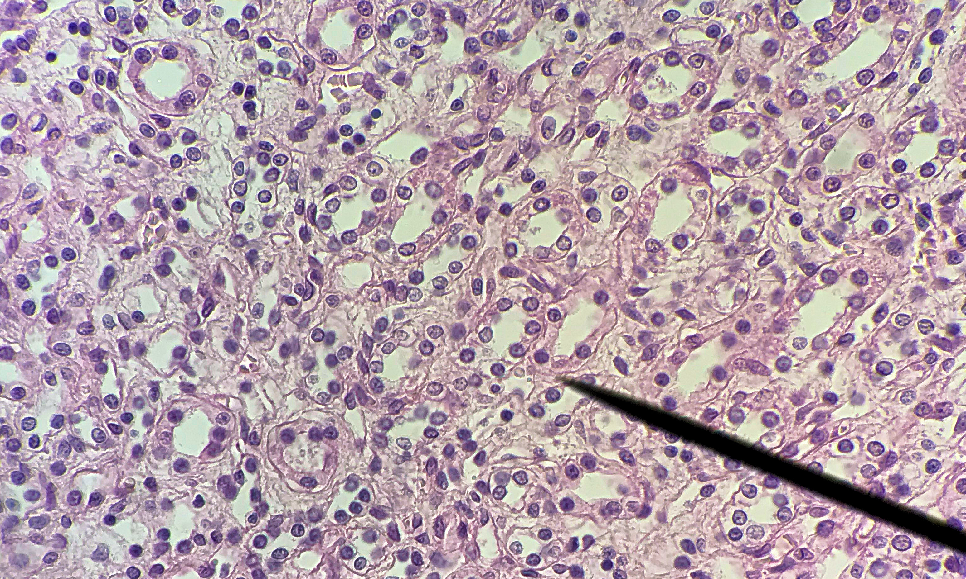
Cells of simple cuboidal epithelium are square or cuboid in shape. Simple cuboidal epithelium is found where absorption or secretion takes place. The cells of simple cuboidal epithelium often are held together by tight junctions so the simple cuboidal cells are the gatekeepers of what can enter or leave that structure. When observed in the kidney, it looks like there are multiple layers of cuboidal cells. However when you look closely, you will see a clear basement membrane at the base of each cell. The tubes of the kidney are arranged so that there are multiple tubes stacked right on top of each other. When observing the slide, see if you can see microvilli, fingerlike projection on cells, in the form of a brush border on the apical side of the cells. The brush border will seem like an uneven surface facing the lumen and it will be hard to focus on the structure. In the kidney slide, you may also be able to spot simple squamosal epithelial found among the tubules. These are capillaries found in the kidney.
Slides on this page were made by students between the spring of 2018 and the spring of 2020. Go through
the diffrent student pictures and compare them to your lab book picture. Then slect one to draw on paper.
Labeled Image
unlabeled Image
1 / 8
2 / 8
3/ 8
4 / 8
5 / 8
6 / 8
7 / 8
8 / 8
❮
❯