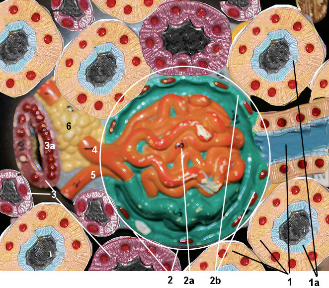
This model shows the histology of the renal cortex. It shows a cross section of a nephron. This view is somewhat
confusing to students. You have to understand that the convoluted tubules go all around the glomerulus. Also notice the difference between the proximal and
distal convoluted tubules. The proximal convoluted tubules have a micro villi that form a brush border. Lastly, try to see that the afferent and efferent arterioles are different
sizes and they house between them the Juxtaglomerular and the macula densa. Find the structures in the word bank on the model and relate it to the histology you
looked at. Then Look at the nephron model and the middle of the kidney section model to see how the nephron really looks. T