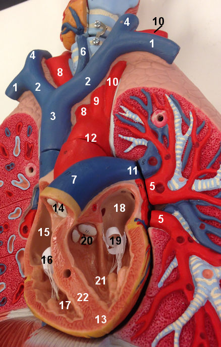
Word Bank
Arteries- Aorta
- Brachiocephalic Artery
- Left Common Carotid Artery
- Left Subclavian Artery
- Pulmonary Artery
- Pulmonary Trunk
Heart
- Aortal semilunar Valve
- Apex
- Bicuspid or Mitral valve
- Interventricular Septum
- Left Atrium
- Left Ventricle
- Pulmonary Semilunar Valve
- Right Atrium
- Right Ventricle
- Tricuspid Valve
- Brachiocephalic vein
- Jugular Vein
- Inferior Thyroid Vein
- Jugular Vein
- Plumonary Veins
- Subclavian vein
- Superior Vena Cava
Notes
- It is jugular vein and carotid artery!
- Veins are bilaterally symmetrical. They are on both sides.
- There is only 1 brachiocephalic artery. This artery leaves the arch of the aorta and splits into the right common carotid and right subclavian artery.
- The left common carotid artery leaves the aortic arch by itself.
- The left subclavian artery leaves the aortic arch by itself.
- Pulmonary arteries are blue on the model because they have oxygen poor blood similar to veins.
- Pulmonary veins are red on the model because they have oxygen rich blood similar to arteries